. 2015 Feb 18;43(3):1937-44.
doi: 10.1093/nar/gkv033. Epub 2015 Jan 27.

An intercalation-locked parallel-stranded DNA tetraplex

Affiliations

An intercalation-locked parallel-stranded DNA tetraplex

Shailesh Tripathi et al. Nucleic Acids Res. .

Abstract

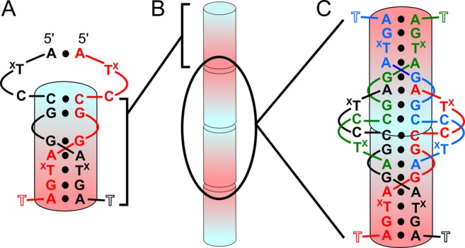
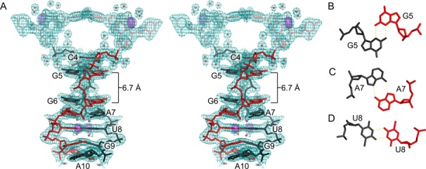
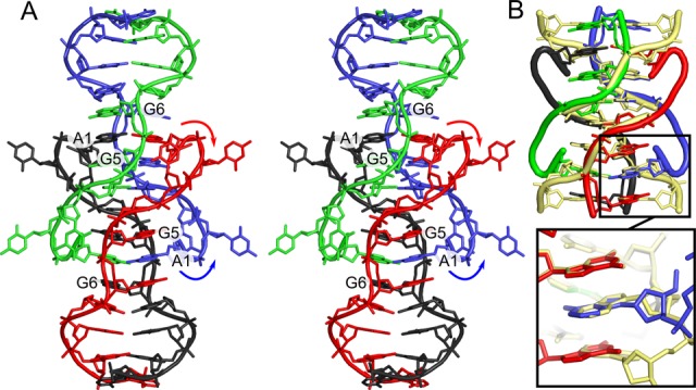
DNA has proved to be an excellent material for nanoscale construction because complementary DNA duplexes are programmable and structurally predictable. However, in the absence of Watson-Crick pairings, DNA can be structurally more diverse. Here, we describe the crystal structures of d(ACTCGGATGAT) and the brominated derivative, d(AC(Br)UCGGA(Br)UGAT). These oligonucleotides form parallel-stranded duplexes with a crystallographically equivalent strand, resulting in the first examples of DNA crystal structures that contains four different symmetric homo base pairs. Two of the parallel-stranded duplexes are coaxially stacked in opposite directions and locked together to form a tetraplex through intercalation of the 5'-most A-A base pairs between adjacent G-G pairs in the partner duplex. The intercalation region is a new type of DNA tertiary structural motif with similarities to the i-motif. (1)H-(1)H nuclear magnetic resonance and native gel electrophoresis confirmed the formation of a parallel-stranded duplex in solution. Finally, we modified specific nucleotide positions and added d(GAY) motifs to oligonucleotides and were readily able to obtain similar crystals. This suggests that this parallel-stranded DNA structure may be useful in the rational design of DNA crystals and nanostructures.


Figures

Figure 1.
Figure 1.
Secondary structure and crystal packing. (A) Secondary structure of interactions formed between two identical DNA strands. The ps duplex region is shown as the shaded cylinder going from 5′ (blue) to 3′ (red). TX represents dT or 5-Br dU residues. T11 was mostly disordered. (B) Representation of coaxial 5′–5′ and 3′–3′ stacking of duplexes along the crystallographic c cell axis. (C) Secondary structure of two duplexes at the 5′–5′ interface with each of the four strands colored differently. The two duplexes stack on the C4–C4 pairs.
Figure 2.
Figure 2.
Parallel-stranded duplex. (A) Stereoview of residues 1–10 from two monomers that form a ps duplex shown with 2mFo–DFc electron density contoured at 0.75 σ. Parallel-stranded duplex residues are labeled. Residues outside the duplex region (A1–U3) are semi-transparent. Anomalous difference electron density contoured at 5 σ (violet surface) corresponds to bromine atoms used for phasing. Water molecules are shown as red spheres. The gap between residues G5 and G6 is 6.7 Å. (B–D) Individual base pairs showing hydrogen bonding between identical residues. (B) N2–N3 sugar edge interactions between G5–G5, G6–G6 and G9–G9 homo base pairs. (C) N6–N7 Hoogsteen interactions between A7–A7 and A10–A10. (D) Symmetric N3–O4 hydrogen bonding observed for U8–U8 homo base pair.
Figure 3.
Figure 3.
5′–5′ stacking. Stereo view of orthogonally stacked C4–C4 homo-base pairs (black, red, blue, green) of two ps homoduplexes. These 5′–5′ stacking interactions are superpositioned with a single step of an i-motif to illustrate the structural similarity (PDB ID: 1CN0; orange).
Figure 4.
Figure 4.
An intercalation-locked tetraplex. (A) Stereo view cartoon and stick representation of a tetraplex with all four strands colored differently. Arrows indicated flipping of A1 residues with respect to ps duplex region. A1–A1 base pairs are intercalated between G5–G5 and G6–G6 pairs of opposite duplex, locking the two duplexes into a tetraplex. (B) Superposition the 5′–5′ interface and A1 residues with 5′–5′ stacked ps CGAA motifs (yellow) (29). C2 and U3 have been removed for clarity. The motifs are structurally similar, and the intercalated A1 residues superpose with first A–A pair of the GA dinucleotide step from the CGAA motif (inset).
Figure 5.
Figure 5.
2D-NOESY NMR spectra. (A) Characteristic C4–C4(+) imino signal at 14.95 ppm and its identified cross peaks. (B) Peak assignments indicating G6–A7 and G9–A10 interstrand stacking in 2D-NOESY NMR spectra, consistent with the ps homo duplex crystal structure. G6 H8 and A7 H2 cross peaks and G9 H8 and A10 H2 are labeled and indicated by intersecting lines. (C) 2D-NOESY NMR spectra indicating interactions between A1 and G5/G6. Cross peaks indicate NOE between these residues, however, peak assignments were not complete for the A1 stack so specific protons have not been assigned for A1.

References

    1. Chen J.H., Seeman N.C. Synthesis from DNA of a molecule with the connectivity of a cube. Nature. 1991;350:631–633. - PubMed
    1. Winfree E., Liu F., Wenzler L.A., Seeman N.C. Design and self-assembly of two-dimensional DNA crystals. Nature. 1998;394:539–544. - PubMed
    1. Goodman R.P., Schaap I.A.T., Tardin C.F., Erben C.M., Berry R.M., Schmidt C.F., Turberfield A.J. Rapid chiral assembly of rigid DNA building blocks for molecular nanofabrication. Science. 2005;310:1661–1665. - PubMed
    1. Rothemund P.W.K. Folding DNA to create nanoscale shapes and patterns. Nature. 2006;440:297–302. - PubMed
    1. Zheng J., Birktoft J.J., Chen Y., Wang T., Sha R., Constantinou P.E., Ginell S.L., Mao C., Seeman N.C. From molecular to macroscopic via the rational design of a self-assembled 3D DNA crystal. Nature. 2009;461:74–77. - PMC - PubMed

Publication types

MeSH terms

LinkOut - more resources