. 2015 Feb 18;43(3):1883-93.
doi: 10.1093/nar/gkv032. Epub 2015 Jan 27.

The principal mRNA nuclear export factor NXF1:NXT1 forms a symmetric binding platform that facilitates export of retroviral CTE-RNA

Affiliations

The principal mRNA nuclear export factor NXF1:NXT1 forms a symmetric binding platform that facilitates export of retroviral CTE-RNA

Shintaro Aibara et al. Nucleic Acids Res. .

Abstract

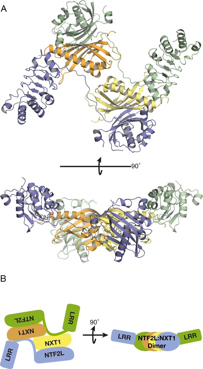
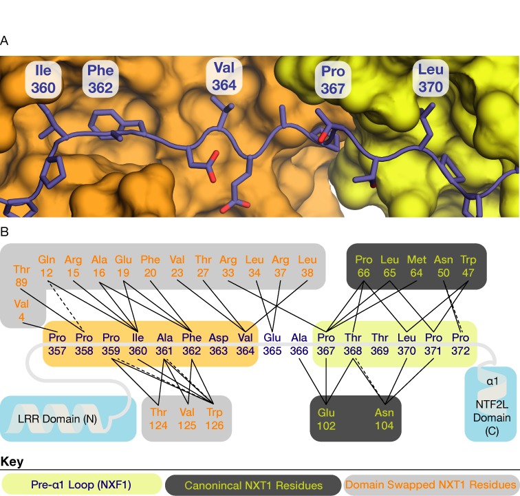
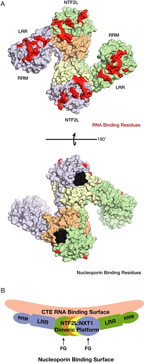
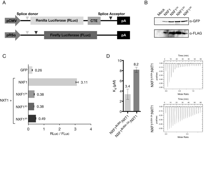
The NXF1:NXT1 complex (also known as TAP:p15) is a general mRNA nuclear export factor that is conserved from yeast to humans. NXF1 is a modular protein constructed from four domains (RRM, LRR, NTF2-like and UBA domains). It is currently unclear how NXF1:NXT1 binds transcripts and whether there is higher organization of the NXF1 domains. We report here the 3.4 Å resolution crystal structure of the first three domains of human NXF1 together with NXT1 that has two copies of the complex in the asymmetric unit arranged to form an intimate domain-swapped dimer. In this dimer, the linkers between the NXF1 LRR and NTF2-like domains interact with NXT1, generating a 2-fold symmetric platform in which the RNA-binding RRM, LRR and NTF2-like domains are arranged on one face. In addition to bulk transcripts, NXF1:NXT1 also facilitates the export of unspliced retroviral genomic RNA from simple type-D retroviruses such as SRV-1 that contain a constitutive transport element (CTE), a cis-acting 2-fold symmetric RNA stem-loop motif. Complementary structural, biochemical and cellular techniques indicated that the formation of a symmetric RNA binding platform generated by dimerization of NXF1:NXT1 facilitates the recognition of CTE-RNA and promotes its nuclear export.


Figures

Figure 1.
Figure 1.
(A) Overview of the 3.4 Å resolution crystal structure of NXF1ΔNΔUBA:NXT1 and (B) Highly schematic illustration of the arrangement of domains in the domain-swapped dimer. Two copies of the protein were present in the asymmetric unit and continuous density was observed between the LRR and NTF2L domains. Density for the RRM domain was not observed, probably as a result of the flexibility of the linker between the RRM and LRR domain. The two copies of the protein in the asymmetric unit of the crystal formed a domain-swapped dimer where the domains were placed roughly in the same plane to form a flat platform with pseudo-2-fold symmetry.
Figure 2.
Figure 2.
(A) Detailed view of the interaction between the linker between the LRR and NTF2L domains (blue) and the two copies of NXT1 (orange and yellow) in the asymmetric unit. Ile360, Phe362, and Val364 made extensive hydrophobic contacts with a domain-swapped NXT1 (orange) in a non-canonical manner. Interactions between the pre-α1 loop and the canonical copy of NXT1 (yellow) that was previously observed in the single domain structure (PDB ID: 1JKG) were also conserved in this multi-domain structure. (B) Schematic representation of the interactions between the LRR–NTF2L linker and the two copies of NXT1. Solid lines represent hydrophobic interactions and dotted lines represent putative hydrogen bonds. Key: pre-α1 loop residues (blue text on yellow); canonical NXT1 residues (yellow text on dark grey); domain-swapped NXT1 residues (orange text on light gray).
Figure 3.
Figure 3.
(A) Structural model in which two copies of NXF1RRM-LRR:CTE-B (PDB ID: 3RW6) were superimposed onto the two LRR domain present in the dimeric structure. The two ends of the stem loop of CTE-B face each other. (B) The ab initio reconstruction obtained from SAXS data of in vitro transcribed full-length CTE-RNA. The overall length of the full-length CTE-RNA was consistent with an elongated stem loop compatible with a length of 160 Å that is comparable with the model proposed in panel A in which the RNA was approximately 140 Å long. (C) Schematic representation of the model shown in panel A where the two CTE-B modules are orientated in a 2-fold symmetric manner. This arrangement of the two CTE-B roughly correspond to a full-length CTE-RNA which has 2-fold symmetry in the center that could span across the NXF1:NXT1 dimer.
Figure 4.
Figure 4.
(A) Surface representation of the protein component from the model shown in Figure 3A. Residues implicated in RNA binding have been colored in red, whereas residues involved in contacts with nucleoporins have been colored in black. Residues not implicated in either RNA or nucleoporin binding have been colored according to the schematics used in Figure 3C. (B) Side view of the schematic of the model proposed for CTE-RNA binding. Importantly the FG nucleoporin binding sites are placed in a structurally compatible location with respect to the proposed RNA binding surface.
Figure 5.
Figure 5.
(A) Western blot analysis demonstrating the effective knock-down of NXF1 by siRNA silencing. The GFP-NXF1R-3A, GFP-NXF1R-3A and GFP-NXF1R-3A mutants had a silent mutation that resisted siRNA-based degradation. (B) Recombinant expression and purification of the mutant NXF1 constructs demonstrate that the NXT1 binding ability of the three mutant NXF1 was not altered. (C) Confocal microscopy demonstrates that cells expressing the three GFP-labeled NXF1 mutants that contained a silent mutation to resist siRNA-based degradation (GFP-NXF1R-3A, GFP-NXF1R-3A and GFP-NXF1R-3A) were able to rescue the accumulation of poly(A)+ RNA in the nucleus, whereas cells in which GFP fluorescence was not present showed nuclear accumulation of poly(A)+ RNA.
Figure 6.
Figure 6.
(A) Schematic of the two plasmids used in the modified dual luciferase reporter assay. The gene for RLuc is fused with the CTE, and thus in the presence of functional NXF1:NXT1 is expressed at higher levels than FLuc. (B) Western blot analysis confirmed that the triple mutations in the LRR–NTF2L linker did not alter levels of expression in HEK293F cells. Probing for FLAG-NXT1 also confirmed that all three NXF1 mutants (GFP-NXF1R-3A, GFP-NXF1R-3A and GFP-NXF1R-3A) retained binding to NXT1. (C) Although wild-type NXF1 generated a 12-fold increase in the RLuc signal relative to that seen with a GFP control, all three NXF1 mutants tested showed greatly reduced enhancement of the RLuc signal. (D) ITC also showed that the triple alanine mutant (NXF13A) has a ∼2.5-fold reduction in binding affinity to full CTE-RNA compared to the wild-type protein.

References

    1. Niño C.A., Hérissant L, Babour A., Dargemont C. mRNA nuclear export in yeast. Chem. Rev. 2013;113:8523–8545. - PubMed
    1. Natalizio B.J., Wente S.R. Postage for the messenger: designating routes for nuclear mRNA export. Trends Cell Biol. 2013;23:365–373. - PMC - PubMed
    1. Stewart M. Nuclear export of mRNA. Trends Biochem. Sci. 2010;35:609–617. - PubMed
    1. Santos-Rosa H., Moreno H., Simos G., Segref A., Fahrenkrog B., Panté N., Hurt E. Nuclear mRNA export requires complex formation between Mex67p and Mtr2p at the nuclear pores. Mol. Cell. Biol. 1998;18:6826–6838. - PMC - PubMed
    1. Grüter P., Tabernero C., von Kobbe C., Schmitt C., Saavedra C., Bachi A., Wilm M., Felber B.K., Izaurralde E. TAP, the human homolog of Mex67p, mediates CTE-dependent RNA export from the nucleus. Mol. Cell. 1998;1:649–659. - PubMed

Publication types