. 2015 Apr 30;125(18):2798-805.
doi: 10.1182/blood-2014-07-590398. Epub 2015 Jan 27.

Interleukin-13 is overexpressed in cutaneous T-cell lymphoma cells and regulates their proliferation

Affiliations

Interleukin-13 is overexpressed in cutaneous T-cell lymphoma cells and regulates their proliferation

Larisa J Geskin et al. Blood. .

Abstract

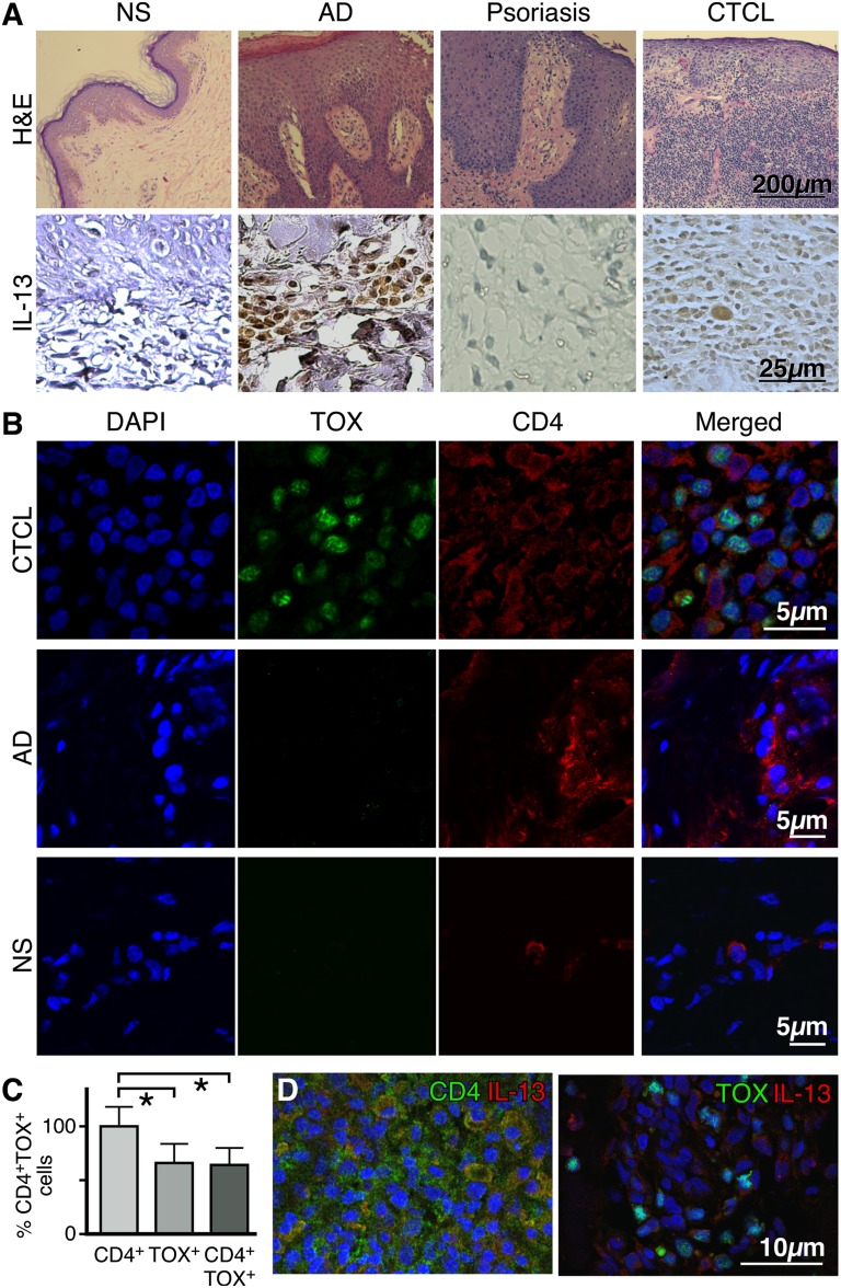
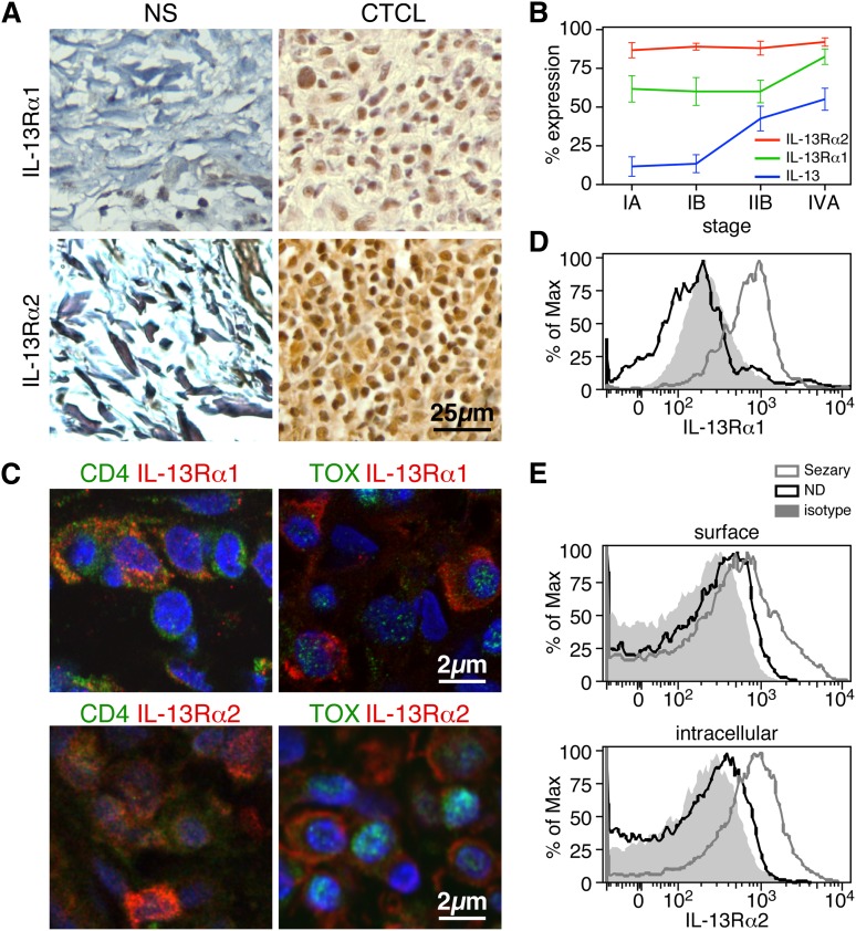
Cutaneous T-cell lymphomas (CTCLs) primarily affect skin and are characterized by proliferation of mature CD4(+) T-helper cells. The pattern of cytokine production in the skin and blood is considered to be of major importance for the pathogenesis of CTCLs. Abnormal cytokine expression in CTCLs may be responsible for enhanced proliferation of the malignant cells and/or depression of the antitumor immune response. Here we show that interleukin-13 (IL-13) and its receptors IL-13Rα1 and IL-13Rα2 are highly expressed in the clinically involved skin of CTCL patients. We also show that malignant lymphoma cells, identified by the coexpression of CD4 and TOX (thymus high-mobility group box), in the skin and blood of CTCL patients produce IL-13 and express both receptors. IL-13 induces CTCL cell growth in vitro and signaling through the IL-13Rα1. Furthermore, antibody-mediated neutralization of IL-13 or soluble IL-13Rα2 molecules can lead to inhibition of tumor-cell proliferation, implicating IL-13 as an autocrine factor in CTCL. Importantly, we established that IL-13 synergizes with IL-4 in inhibiting CTCL cell growth and that blocking the IL-4/IL-13 signaling pathway completely reverses tumor-cell proliferation. We conclude that IL-13 and its signaling mediators are novel markers of CTCL malignancy and potential therapeutic targets for intervention.


Figures

Figure 1
Figure 1
IL-13 is expressed by tumor cells in the skin lesions of CTCL patients. (A, upper panel) Representative hematoxylin and eosin stains of normal skin (NS), atopic dermatitis (AD), psoriasis, and CTCL skin (original magnification ×100). (A, lower panel) Immunohistochemical staining for IL-13 expression in skin biopsies from NS (n = 3), AD (n = 5), psoriasis (n = 5), or CTCL (n = 17) (original magnification ×400). (B) Double-color immunofluorescence labeling of frozen skin samples from NS (n = 3), AD (n = 3), and CTCL (n = 7) biopsies stained with antibodies to CD4 (surface staining) and TOX (nuclear staining). A representative example is shown (original magnification ×1000). (C) Proportion of CD4+ and TOX+ cells in the skin of CTCL patients. Error bars are mean ± standard deviation (SD) (n = 7). Statistics were derived by Student t test. (D) Representative example of double-color immunofluorescence staining for CD4 and IL-13 (upper panel) or TOX and IL-13 (lower panel) (original magnification ×400). Skin samples from 7 CTCL patients were analyzed giving similar results. 4,6 diamidino-2-phenylindole stains nuclei.
Figure 2
Figure 2
IL-13 receptors are expressed by tumor cells in the skin lesions of CTCL patients and in the peripheral blood CD4+ T cells of Sézary patients. (A) Expression by immunohistochemistry of IL-13Rα1 (upper panel) and IL-13Rα2 (lower panel) in biopsies from CTCL and normal skin (original magnification ×400). Representative examples are shown (n = 17 CTCL, n = 3 NS). (B) The percentages of IL-13+, IL-13Rα1+, and IL-13Rα2+ cells in CTCL skin biopsies at different disease stages are shown. Error bars are mean ± SD. (C) Representative examples of IL-13Rα1 or IL-13Rα2 costaining with CD4 or TOX by immunofluorescence of frozen CTCL skin biopsies (original magnification ×1000, n = 7). Flow cytometry expression of IL-13Rα1 (D) or IL-13Rα2 (E) in CD4+ T cells from the peripheral blood of SS patients. Representative examples out of 5 SS patients tested.
Figure 3
Figure 3
Blocking the IL-13–signaling pathway inhibits SS CD4+ T-cell proliferation. (A) Freshly isolated CD4+ T cells from SS patients were cultured in vitro for 5 days and treated with 10, 50, 100, or 500 ng/mL of IL-13. Proliferation was determined by MTT assay and data are depicted as means ± SD compared with untreated cells. Statistics were gathered by ANOVA followed by post hoc Dunnett test. (B) CD4+ T cells from SS patients (upper panel) or NDs (lower panel) were treated for 15 minutes with IL-4 or IL-13 and pSTAT-6 was determined by intracellular staining as described in Material and methods. Shown is a representative example of 5 independent experiments giving similar results. (C) Immunohistochemical analysis of pSTAT-6 from NS (n = 3), AD skin (n = 3), and stage I (n = 4) and stage IV (n = 4) CTCL skin biopsies. Representative examples are shown (original magnification ×400). (D) The percentages of pSTAT-6+ cells after quantification of 20 HPF for each sample are shown. Error bars are mean ± SD. Statistics were gathered by ANOVA followed by post hoc Tukey test. (E) Effect of a soluble IL-13Rα2 (2.5 μg/mL), an anti-IL-13 antibody (6.5 μg/mL), an anti-IL-4 antibody (10 μg/mL), or a pSTAT-6 inhibitor (AS1517499, 100 nM) on SS CD4+ T-cell proliferation. The proliferation inhibition rate was detected by MTT assay after 5 days in culture; the data are shown as means ± SD; ***P < .001 compared with the untreated cells. Statistics were gathered by ANOVA followed by post hoc Tukey test.

Comment in

Similar articles

Cited by

References

    1. Samuelson E. Cutaneous T-cell lymphomas. Semin Oncol Nurs. 1998;14(4):293–301. - PubMed
    1. Diamandidou E, Cohen PR, Kurzrock R. Mycosis fungoides and Sezary syndrome. Blood. 1996;88(7):2385–2409. - PubMed
    1. Sterry W, Mielke V. CD4+ cutaneous T-cell lymphomas show the phenotype of helper/inducer T cells (CD45RA-, CDw29+). J Invest Dermatol. 1989;93(3):413–416. - PubMed
    1. Zackheim HS, Amin S, Kashani-Sabet M, McMillan A. Prognosis in cutaneous T-cell lymphoma by skin stage: long-term survival in 489 patients. J Am Acad Dermatol. 1999;40(3):418–425. - PubMed
    1. Diamandidou E, Colome M, Fayad L, Duvic M, Kurzrock R. Prognostic factor analysis in mycosis fungoides/Sézary syndrome. J Am Acad Dermatol. 1999;40(6 Pt 1):914–924. - PubMed

Publication types