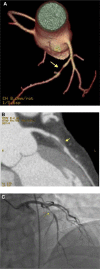
Multi-Detector Coronary CT Imaging for the Identification of Coronary Artery Stenoses in a "Real-World" Population
- PMID: 25628513
- PMCID: PMC4284987
- DOI: 10.4137/CMC.S18223
Multi-Detector Coronary CT Imaging for the Identification of Coronary Artery Stenoses in a "Real-World" Population
Abstract
Background: Multi-detector computed tomography (CT) has emerged as a modality for the non-invasive assessment of coronary artery disease (CAD). Prior studies have selected patients for evaluation and have excluded many of the "real-world" patients commonly encountered in daily practice. We compared 64-detector-CT (64-CT) to conventional coronary angiography (CA) to investigate the accuracy of 64-CT in determining significant coronary stenoses in a "real-world" clinical population.
Methods: A total of 1,818 consecutive patients referred for 64-CT were evaluated. CT angiography was performed using the GE LightSpeed VCT (GE(®) Healthcare). Forty-one patients in whom 64-CT results prompted CA investigation were further evaluated, and results of the two diagnostic modalities were compared.
Results: A total of 164 coronary arteries and 410 coronary segments were evaluated in 41 patients (30 men, 11 women, age 39-85 years) who were identified by 64-CT to have significant coronary stenoses and who thereafter underwent CA. The overall per-vessel sensitivity, specificity, positive predictive value, negative predictive value, and accuracy at the 50% stenosis level were 86%, 84%, 65%, 95%, and 85%, respectively, and 77%, 93%, 61%, 97%, and 91%, respectively, in the per-segment analysis at the 50% stenosis level.
Conclusion: 64-CT is an accurate imaging tool that allows a non-invasive assessment of significant CAD with a high diagnostic accuracy in a "real-world" population of patients. The sensitivity and specificity that we noted are not as high as those in prior reports, but we evaluated a population of patients that is typically encountered in clinical practice and therefore see more "real-world" results.
Keywords: 64-detector coronary computed tomography; coronary angiography; coronary artery disease.
Figures


Similar articles
-
Significant coronary artery stenosis: comparison on per-patient and per-vessel or per-segment basis at 64-section CT angiography.Radiology. 2007 Jul;244(1):112-20. doi: 10.1148/radiol.2441060332. Radiology. 2007. PMID: 17581898
-
Heart imaging: the accuracy of the 64-MSCT in the detection of coronary artery disease.Eur Rev Med Pharmacol Sci. 2009 May-Jun;13(3):163-71. Eur Rev Med Pharmacol Sci. 2009. PMID: 19673166
-
16-detector row computed tomographic coronary angiography in patients undergoing evaluation for aortic valve replacement: comparison with catheter angiography.Clin Radiol. 2006 Sep;61(9):749-57. doi: 10.1016/j.crad.2006.04.016. Clin Radiol. 2006. PMID: 16905381
-
Clinical value of 16-slice multi-detector CT compared to invasive coronary angiography.Int J Cardiovasc Intervent. 2005;7(1):21-8. doi: 10.1080/14628840510011207. Int J Cardiovasc Intervent. 2005. PMID: 16019611
-
Coronary artery stenosis: direct comparison of four-section multi-detector row CT and 3D navigator MR imaging for detection--initial results.Radiology. 2005 Jan;234(1):98-108. doi: 10.1148/radiol.2341031325. Epub 2004 Nov 18. Radiology. 2005. PMID: 15550371
Cited by
-
Cardiovascular Imaging: Current Developments in Research and Clinical Practice.Clin Med Insights Cardiol. 2016 Apr 3;8(Suppl 4):57-61. doi: 10.4137/CMC.S38846. eCollection 2014. Clin Med Insights Cardiol. 2016. PMID: 27081320 Free PMC article. No abstract available.
References
-
- Raff G, Gallagher MJ, O’Neill WW, Goldstein JA. Diagnostic accuracy of noninvasive coronary angiography using 64-slice spiral computed tomography. J Am Coll Cardiol. 2005;46:552–7. - PubMed
-
- Mollet NR, Cademartiri F, van Mieghem CA, et al. High-resolution spiral computed tomography coronary angiography in patients referred for diagnostic conventional coronary angiography. Circulation. 2005;112:2318–23. - PubMed
-
- Ropers D, Baum U, Pohle K, et al. Detection of coronary artery stenoses with thin-slice multi-detector row spiral computed tomography and multiplanar reconstruction. Circulation. 2003;107:664–6. - PubMed
-
- Kuettner A, Trabold T, Schroeder S, et al. Noninvasive detection of coronary lesions using 16-detector multislice spiral computed tomography technology: initial clinical results. J Am Coll Cardiol. 2004;44:1230–7. - PubMed
-
- Achenbach S, Giesler T, Ropers D, et al. Detection of coronary artery stenoses by contrast-enhanced, retrospectively electrocardiographically gated, multislice spiral computed tomography. Circulation. 2001;103:2535–8. - PubMed
LinkOut - more resources
Full Text Sources
Other Literature Sources
Miscellaneous

