Review
. 2015 Jan 12:8:450.
doi: 10.3389/fncel.2014.00450. eCollection 2014.

Cellular commitment in the developing cerebellum

Affiliations
Review

Cellular commitment in the developing cerebellum

Hassan Marzban et al. Front Cell Neurosci. .

Abstract

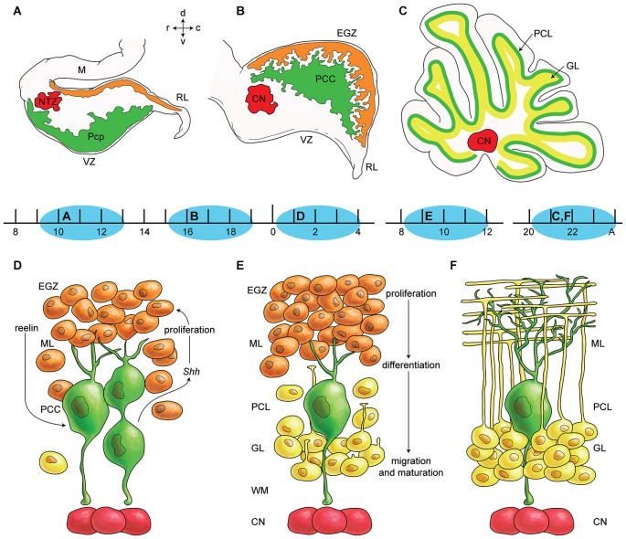
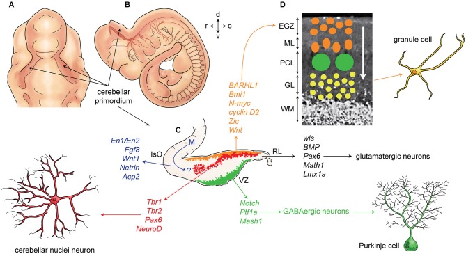
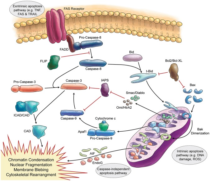
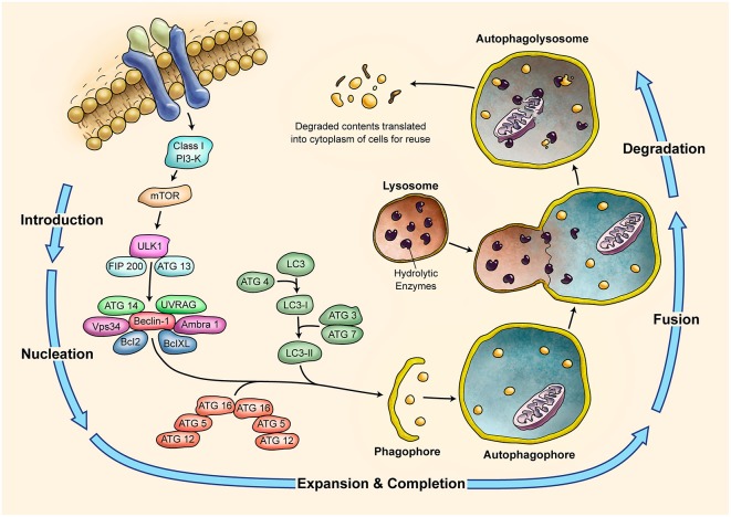
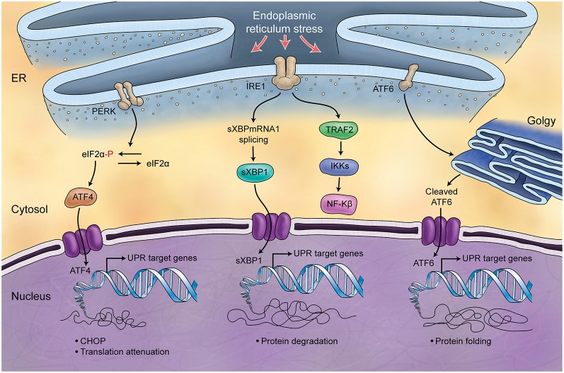
The mammalian cerebellum is located in the posterior cranial fossa and is critical for motor coordination and non-motor functions including cognitive and emotional processes. The anatomical structure of cerebellum is distinct with a three-layered cortex. During development, neurogenesis and fate decisions of cerebellar primordium cells are orchestrated through tightly controlled molecular events involving multiple genetic pathways. In this review, we will highlight the anatomical structure of human and mouse cerebellum, the cellular composition of developing cerebellum, and the underlying gene expression programs involved in cell fate commitments in the cerebellum. A critical evaluation of the cell death literature suggests that apoptosis occurs in ~5% of cerebellar cells, most shortly after mitosis. Apoptosis and cellular autophagy likely play significant roles in cerebellar development, we provide a comprehensive discussion of their role in cerebellar development and organization. We also address the possible function of unfolded protein response in regulation of cerebellar neurogenesis. We discuss recent advancements in understanding the epigenetic signature of cerebellar compartments and possible connections between DNA methylation, microRNAs and cerebellar neurodegeneration. Finally, we discuss genetic diseases associated with cerebellar dysfunction and their role in the aging cerebellum.

Keywords: DNA methylation; aging; apoptosis; autophagy; brain development; cerebellum structure; epigenetics.


Figures

Figure 1
Figure 1
Germinal zones in the developing cerebellum. (A–C) Schematic illustration of the spatiotemporal parameters at sagittal sections of the early cerebellar development (embryonic (E) day 10–13 (E10–E13) (A), E16–E17 (B), and postnatal (P) day 20 (P20) to adult (C). Neuroepithelium of 4th ventricle (ventricular zone (VZ)) is sources of all GABAergic neurons including Purkinje cells (green), Rhombic lip (RL) is sources of glutamatergic neurons including cerebellar nuclei neurons (red) and external germinal zone (orange; source of granule cells). (D–F) Schematic illustrations of the spatiotemporal parameters in corticogensis in which Purkinje cells cluster disperse in the monolayer and granular layer form. A cartoon of cerebellar cortex at around P4 (D), at around P10 (E), and in adult (F) is shown. Purkinje cells (green) express SHH that increases proliferative activity of external germinal zone (EGZ) cells (precursor of granule cells). Reelin express from precursor of granule cells and causes dispersal of Purkinje cells cluster (D) to monolayer (E–F). Granule cells differentiate and migrate cross Purkinje cells layer to final destination i.e., granular layer and granule cells development is completed by maturation in this layer. Abbreviations: Pcc: Purkinje cell clusters, Purkinje cells precursor: pcp, mesencephalon: m, rhombic lip: RL, E: embryonic day, EGZ: external germinal layer (zone), gc: granule cells, m: mesencephalon, NTZ: nuclear transitory zone, A: Adult, pcl: Purkinje cell layer, RL: rhombic lip, ml: molecular layer.
Figure 2
Figure 2
Development of cerebellum and corticogenesis. (A–B) Dorsal and lateral aspect of the mouse embryo at embryonic day E10-E11, showing outline of cerebellar primordium (indicated by arrows) and mesencephalon (midbrain) (m). (C) Schematic illustration of the developing cerebellum at about E11 indicates ventricular germinal zone (green) and genes are involved in neurogenesis of GABAergic neurons such as Purkinje cells. Rhombic lip is germinal zone that under control of genes such as Wls, Bmp, math1, pax6, and Lmx1a generate almost all glutamatergic neurons in cerebellum. Rhombic lip derived produce cerebellar nuclei neurons (red), and external germinal zones precursors (orange). An arrow from mesencephalon (m) indicates a germinal zone for group of cells derived from mesencephalon to the cerebellar primordium. (D) A section of cerebellum at around P4 indicate external germinal zone (EGZ) that after proliferation migrate through the Purkinje cells to the granular layer that is location of granule cells. Abbreviation; Iso: isthmic organizer, m: mesencephalon, EGZ: external germinal zone, ML: molecular layer, PCL: Purkinje cell layer, GL: granular layer, WM: white matter, 4thV: fourth ventricle, r: rostral, c: caudal, d: dorsal, v: ventral.
Figure 3
Figure 3
Schematic Representation of Apoptosis. In general apoptosis divides into extrinsic and intrinsic pathway. Death receptors (like FAS) are involved in extrinsic pathway, which later can activate caspase-8. Caspase-8 activates caspase-3 in two separate ways (direct activation or activation via caspase-9). Stress signals and DNA damage triggers intrinsic apoptosis pathway via mitochondria. Intrinsic apoptosis (mitochondrial apoptosis) is divided to caspase-dependent or caspase-independent pathways.
Figure 4
Figure 4
Schematic Representation of Autophagy Pathway. Autophagy is recognized as a major tool to degrade damaged organelles and misfolded proteins via lysosomal pathway. Autophagy is an active flux which includes five different steps; introduction or initiation, phagosome nucleation, phagosome expansion and completion, phagosome and lysosme fusion (autophagolysosome formation), and finally degradation. It is a tightly regulated mechanism and several ATG molecules are included in its regulation.
Figure 5
Figure 5
Schematic Representation of Unfolded Protein Response. Endoplasmic reticulum (ER) is involved in the processing of proteins and is responsible for regulation of protein folding. ER chaperones (PERK, IRE1, ATF6) are deactivated in normal conditions while in stress conditions and increase of misfolded proteins in ER, they will be activated (UPR) and differently control protein biosynthesis, cell survival, protein translation and cell cycle.

Similar articles

Cited by

References

    1. Abrahám H., Tornóczky T., Kosztolányi G., Seress L. (2001). Cell formation in the cortical layers of the developing human cerebellum. Int. J. Dev. Neurosci. 19, 53–62. 10.1016/s0736-5748(00)00065-4 - DOI - PubMed
    1. Aguilar A., Meunier A., Strehl L., Martinovic J., Bonniere M., Attie-Bitach T., et al. . (2012). Analysis of human samples reveals impaired SHH-dependent cerebellar development in Joubert syndrome/Meckel syndrome. Proc. Natl. Acad. Sci. U S A 109, 16951–16956. 10.1073/pnas.1201408109 - DOI - PMC - PubMed
    1. Alder J., Lee K. J., Jessell T. M., Hatten M. E. (1999). Generation of cerebellar granule neurons in vivo by transplantation of BMP-treated neural progenitor cells. Nat. Neurosci. 2, 535–540. 10.1038/9189 - DOI - PubMed
    1. Altman J., Bayer S. A. (1985a). Embryonic development of the rat cerebellum. I. Delineation of the cerebellar primordium and early cell movements. J. Comp. Neurol. 231, 1–26. 10.1002/cne.902310103 - DOI - PubMed
    1. Altman J., Bayer S. A. (1985b). Embryonic development of the rat cerebellum. II. Translocation and regional distribution of the deep neurons. J. Comp. Neurol. 231, 27–41. 10.1002/cne.902310104 - DOI - PubMed