

Genomic instability in complicated and uncomplicated Egyptian schistosomiasis haematobium patients
- PMID: 25628757
- PMCID: PMC4307227
- DOI: 10.1186/s13039-014-0104-5
Genomic instability in complicated and uncomplicated Egyptian schistosomiasis haematobium patients
Abstract
Background: Exploration of genetic changes during active Schistosoma infection is important for anticipation and prevention of chronic sequelae. This study aimed to explore the genomic instability in chromosomal and cellular kinetics in Egyptians suffering from uncomplicated active schistosomiasis haematobium infection in addition to chronic schistosomiasis haematobium cases complicated by bilharzial-associated bladder cancer (BAC).
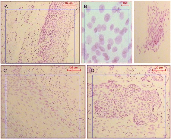
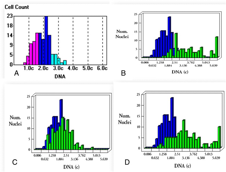
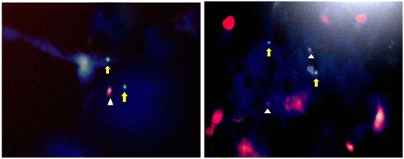
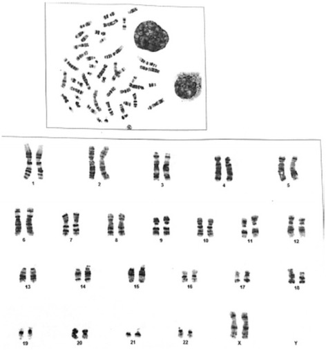
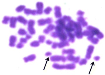
Results: This study was conducted on 46 schistosomiasis haemotobium cases, 22 were active (Viable S. haematobium eggs in urine samples as detected by microscopy) and 24 were chronic complicated with bladder cancer. Three cytogenetic techniques were applied; the first was quantitative nuclear-morphocytometry by means of which the Feulgen-stained nuclei were analyzed for parameters including shape, size, integrated optical-density and nuclear area. The second was Fluorescent In-Situ Hybridization (FISH) for specific p53gene-locus of chromosome 17 and the third technique was karyotyping. Concerning chronic complicated cases, the mean ± SD of DNA-content in urinary bladder tissue sections was 3.18 ± 0.65. Five samples (20.83%) of bladder tissue sections of chronic complicated cases showed diploid nuclei, 6 urinary bladder tissue samples (25%) were tetraploid, while 13 bladder samples (54.16%) were aneuploid. Epithelial cells of urine samples demonstrated aneuploidy (mean ± SD = 3.74 ± 0.36).Nuclear contents showed high proliferative DNA index in all urinary epithelial cells. In the acute uncomplicated group, nuclear-DNA of urinary epithelial cells was found diploid with mean nuclear-DNA content of 2.2 ± 0.16SD. Half of these diploid smears had a high proliferation index. The difference between nuclear DNA-contents in acute and chronic cases was significant (P = 0.0001). FISH technique for specific p53gene-locus and karyotyping were done on urinary bladder tissue specimens and peripheral blood monocytes of 8 chronic cases respectively. Three samples (37.5%) with invasive BAC had a deletion of the p53 gene. Karyotyping showed three cases out of the 8 chronic schistosomiasis haematobium patients with chromosomal fragmentations.
Conclusions: DNA morphometry was valuable in detection of gross genetic changes in urothelial tissues. It is an important prognostic factor in established schistosomiasis haematobium induced bladder malignancy. It has the great advantage of being applicable on urine cells making it suitable for the prediction of a tendency towards genetic instability in active schistosomiasis haematobium patients.
Keywords: Chromosomal abnormalities; FISH; Karyotyping; Morphocytometry; Schistosomiasis haematobium.
Figures
References
-
- Mohkatar N, Gouda I, Adel I. Cancer pathology registry (2003–2007), 2nd edn. Cairo, Egypt: The National Cancer Institute at Cairo University.2007
LinkOut - more resources
Full Text Sources
Other Literature Sources
Medical
Research Materials
Miscellaneous
