

Review
doi: 10.4329/wjr.v7.i1.17.
Fetal brain tumors: Prenatal diagnosis by ultrasound and magnetic resonance imaging
Affiliations
- PMID: 25628801
- PMCID: PMC4295174
- DOI: 10.4329/wjr.v7.i1.17
Item in Clipboard
Review
Fetal brain tumors: Prenatal diagnosis by ultrasound and magnetic resonance imaging
World J Radiol.
.
Abstract
Congenital central nervous system tumors diagnosed during pregnancy are rare, and often have a poor prognosis. The most frequent type is the teratoma. Use of ultrasound and magnetic resonance image allows the suspicion of brain tumors during pregnancy. However, the definitive diagnosis is only confirmed after birth by histology. The purpose of this mini-review article is to describe the general clinical aspects of intracranial tumors and describe the main fetal brain tumors.
Keywords: Brain tumors; Fetus; Magnetic resonance imaging; Teratoma; Ultrasound.
Figures
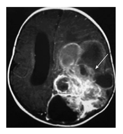
Case of anaplastic astrocytoma. Magnetic resonance imaging (T1 Fast Spin Echo, axial plane) 7 d after birth showing large parieto-occipital lesion with solid and cystic components (white arrow).

Fetus at 15 wk of gestation. Magnetic resonance imaging (T2 haste), (A) coronal plane, and (B) axial plane, showing a large lesion with solid and cystic components involving the whole brain (white arrows), and a severe macrocrania (cranial biometry for 30 wk). Histology confirmed immature teratoma.

Teratoma. A: Ultrasound (US) at 29 wk of gestation (median sagittal plane) showing heterogeneous and hyperechogenic lesion in the suprasellar region (white arrow); B: Fetal magnetic resonance imaging (T2 haste, sagittal plane) at 29 wk of gestation confirming aspects observed on US (white arrow); C: Magnetic resonance imaging (T1 Fast Spin Echo, sagittal plane) 3 d after birth showing suprasellar lesion with solid and cystic components distorting brain anatomy (white arrow). Histology confirmed teratoma.

Craniopharyngioma. (A) Fetal magnetic resonance imaging (T2 haste, sagittal plane) and (B) ultrasound (axial plane) at 28 wk of gestation showing a heterogeneous and hyperechogenic suprasellar lesion and hydrocephalus (white arrows). Histology confirmed craniopharyngioma.
References
-
- Cavalheiro S, Moron AF, Hisaba W, Dastoli P, Silva NS. Fetal brain tumors. Childs Nerv Syst. 2003;19:529–536. - PubMed
-
- Meizner I. Tumors of the Brain. In: Ultrasonography of the prenatal brain., editor. 3rd ed. McGrawHill: New York; 2012. pp. 393–406.
-
- Cassart M, Bosson N, Garel C, Eurin D, Avni F. Fetal intracranial tumors: a review of 27 cases. Eur Radiol. 2008;18:2060–2066. - PubMed
-
- Hoff NR, Mackay IM. Prenatal ultrasound diagnosis of intracranial teratoma. J Clin Ultrasound. 1980;8:247–249. - PubMed
Publication types
LinkOut - more resources
Full Text Sources
Other Literature Sources
