Clinical, endoscopic, and histopathological aspects of sigmoid actinomycosis; a case report and literature review
- PMID: 25628853
- PMCID: PMC4293800
Clinical, endoscopic, and histopathological aspects of sigmoid actinomycosis; a case report and literature review
Abstract
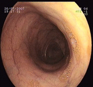
Actinomycosis is a rare and chronic infectious disease caused by a non-spore gram- positive, anaerobic bacterium that rarely infects the colon, in particular the left colon. A 53-year-old woman was referred to us due to chronic abdominal pain, bloating, a few episodes of bloody-mucous rectal discharge, and change of bowel habits. Her medical history and physical examination were unremarkable. Colonoscopy revealed a polypoid mass like lesion located 20 cm proximal to the anal verge above the rectosigmoid junction. Several biopsy samples were taken. Histopathological evaluation showed actinomycosis infection. Consequently the patient was treated with intravenous and then six months oral penicillin. Her complaints and colonic mass resolved totally. Diagnosis of colonic actinomycosis is not an easy task. It is advisable to keep this infection in mind among the differential diagnoses of unusual abdominal masses. Colonoscopy and histopathological examination can be the preferred modality for diagnosis of colonic actinomycosis infection.
Keywords: Actinomycosis; Left colon; Penicillin; Sigmoid.
References
-
- Bennhoff DF. Actinomycosis: diagnostic and therapeutic considerations and a review of 32 cases. Laryngoscope. 1984;94:1198–217. - PubMed
-
- Wong VK, Turmezei TD, Weston VC. Actinomycosis. BMJ. 2011;343:d6099. - PubMed
-
- Naf F, Enzler-Tschudy A, Kuster SP, Uhlig I, Steffen T. Abdominal Actinomycosis Mimicking a Malignant Neoplasm. Surg Infect (Larchmt) 2014;15:462–3. - PubMed
-
- Petrie BA, Schwartz SI, Saltmarsh GF. Intra-abdominal Actinomycosis in Association with Sigmoid Diverticulitis. Am Surg. 2014;80:157–9. - PubMed
Publication types
LinkOut - more resources
Full Text Sources


