. 2015:2015:320642.
doi: 10.1155/2015/320642. Epub 2015 Jan 5.

Impact of viral activators and epigenetic regulators on HIV-1 LTRs containing naturally occurring single nucleotide polymorphisms

Affiliations

Impact of viral activators and epigenetic regulators on HIV-1 LTRs containing naturally occurring single nucleotide polymorphisms

Sonia Shah et al. Biomed Res Int. 2015.

Abstract

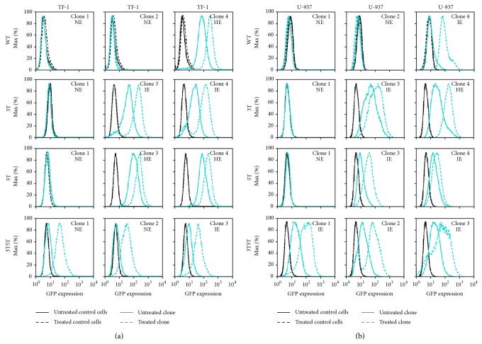
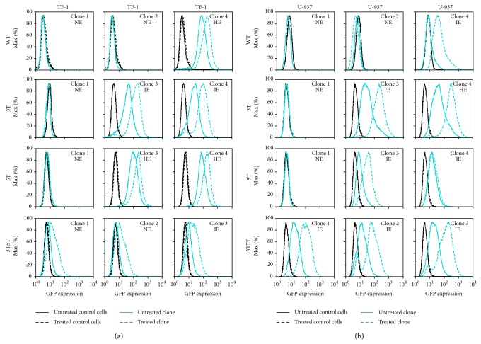
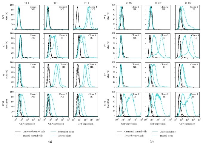
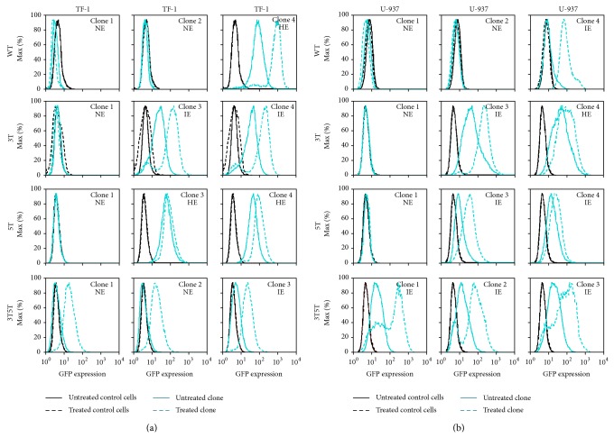
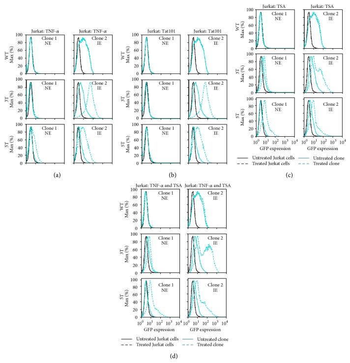
Following human immunodeficiency virus type 1 (HIV-1) integration into host cell DNA, the viral promoter can become transcriptionally silent in the absence of appropriate signals and factors. HIV-1 gene expression is dependent on regulatory elements contained within the long terminal repeat (LTR) that drive the synthesis of viral RNAs and proteins through interaction with multiple host and viral factors. Previous studies identified single nucleotide polymorphisms (SNPs) within CCAAT/enhancer binding protein (C/EBP) site I and Sp site III (3T, C-to-T change at position 3, and 5T, C-to-T change at position 5 of the binding site, respectively, when compared to the consensus B sequence) that are low affinity binding sites and correlate with more advanced stages of HIV-1 disease. Stably transfected cell lines containing the wild type, 3T, 5T, and 3T5T LTRs were developed utilizing bone marrow progenitor, T, and monocytic cell lines to explore the LTR phenotypes associated with these genotypic changes from an integrated chromatin-based microenvironment. Results suggest that in nonexpressing cell clones LTR-driven gene expression occurs in a SNP-specific manner in response to LTR activation or treatment with trichostatin A treatment, indicating a possible cell type and SNP-specific mechanism behind the epigenetic control of LTR activation.


Figures

Figure 1
Figure 1
TNF-α induction of LTR-driven GFP expression is dependent on the basal LTR expression phenotype in hematopoietic progenitor TF-1 cell clones as well as promonocytic U-937 cell clones. Both TF-1 and U-937 cells stably transfected with the LAI LTR (WT, 3T, 5T, and 3T5T) were serially diluted in order to obtain 1 cell in 1 mL of media (approximately one cell in 10 wells of a 96-well plate). Cell clone populations were propagated from the single cell and then were analyzed using flow cytometry for their basal GFP expression. The clonal populations were then designated in one of three categories, nonexpresser (NE), intermediate expresser (IE), and high-expresser (HE), based on their geometric mean fluorescence intensity (MFI) and their percent cell positive values. Each individual cellular clone across all four backbones was treated with TNF-α (20 ng/mL). All experiments were completed in triplicate in three independent experiments. Representative histograms showing levels of GFP expression obtained with the untreated stably transfected cell clone (solid turquoise line) compared to the levels of GFP expression obtained with treated stably transfected cell clone (dashed turquoise line), treated WT TF-1 and U-937 cells (dashed black line), and untreated WT TF-1 and U-937 cells (solid black line) are shown. (a) The NE LAI WT, 3T, and 5T LTR-containing clones could not be induced into driving GFP expression, whereas their GFP-expressing clone counterparts could be induced. All the NE and the IE 3T5T LTR-containing TF-1 clones could be induced to express higher levels of GFP expression. (b) The NE LAI WT, 3T, and 5T LTR-containing clones could not be induced into driving GFP expression, whereas their GFP-expressing clone counterparts could be induced. All of the IE 3T5T LTR-containing clones could be induced to express high levels of GFP.
Figure 2
Figure 2
IL-1β induction of HIV-1 transcription is dependent on basal LTR expression phenotype in TF-1 and U-937 cell clones. TF-1 and U-937 stably transfected cell clones containing the WT, 3T, 5T, or 3T5T LAI LTRs were treated with IL-1β (20 ng/mL) for 24 hours and then analyzed using flow cytometry for their stimulated GFP expression. All experiments were completed in triplicate in three independent experiments. Representative histograms showing levels of GFP expression obtained with the untreated stably transfected cell clone (solid turquoise line), the treated stably transfected cell clone (dashed turquoise line), untreated WT TF-1 and U-937 cells (solid black line), and treated WT TF-1 and U-937 cells (dashed black line) are shown. Designations of nonexpressing (NE), intermediate-expressing (IE), and high-expressing (HE) are determined based on basal levels of LTR-driven gene expression within each clone. (a) LTRs from NE WT, 3T, and 5T TF-1 cells were unable to be induced into driving GFP expression, whereas the cells containing NE 3T5T LTRs were activated following treatment. Additionally, active LTRs could be induced to drive higher levels of GFP expression. (b) LTRs from NE U-937 cells were unable to be induced into driving GFP expression, whereas the cells containing active LTRs could be induced to drive higher levels of GFP expression.
Figure 3
Figure 3
Tat101 transactivation leads to increases in LTR-driven gene transcription within GFP-expressing TF-1 and U-937 cell clones. TF-1 and U-937 stably transfected cell clones containing the WT, 3T, 5T, or 3T5T LAI LTRs were transfected with Tat101 (300 ng) using the Ingenio electroporation solution with an Amaxa nucleofector device and were analyzed for GFP expression using flow cytometry 24 hours after transfection. All experiments were completed in triplicate in three independent experiments. Representative histograms show GFP expression levels from the untreated stably transfected cell clone (solid turquoise line), the treated stably transfected cell clone (dashed turquoise line), untreated WT TF-1 and U-937 cells (solid black line), and treated WT TF-1 and U-937 cells (dashed black line). Within the context of Tat, untreated refers to transfection with the parental pcDNA3.1 plasmid without the Tat gene (in other words, empty vector). Designations of nonexpressing (NE), intermediate-expressing (IE), and high-expressing (HE) are determined based on basal levels of LTR-driven gene expression within each clone. (a) LTRs from NE WT, 3T, and 5T TF-1 cells were unable to be induced into driving GFP expression, whereas LTRs from NE 3T5T TF-1 cells could be induced to drive higher levels of GFP expression. Additionally, the cells containing active LTRs could be induced to drive higher levels of GFP expression. (b) LTRs from NE cells were unable to be induced into driving GFP expression, whereas the cells containing active LTRs could be induced to drive higher levels of GFP expression.
Figure 4
Figure 4
HIV-1 LAI non-GFP-expressing LTR is not inducible by the combination of TSA and TNF-α above levels seen with TNF-α alone in stably transfected TF-1 and U-937 cell clones. Stably transfected TF-1 cell clones containing the WT, 3T, 5T, or 3T5T LAI LTRs were treated with 100 nM trichostatin A (TSA) in combination with TNF-α (20 ng/mL) for 24 hours, along with untreated clone controls as well as untreated and treated TF-1 cells (a) and U-937 cells (b). After 24 hours, cells were analyzed for GFP expression using flow cytometry. All experiments were completed in triplicate in three independent experiments. Representative histograms show levels of GFP expression of the untreated cells (solid black line) compared to the treated cells (dashed black line), and untreated stably transfected cell clones (solid turquoise line) compared to the treated cell clones (dashed turquoise line). TSA alone did not result in any alterations in gene expression within any of the cell clones examined.
Figure 5
Figure 5
LTR-driven gene expression is induced in non-GFP-expressing LAI LTR clones stimulated with various treatments in T-cell clones. Jurkat cells stably transfected with the LAI LTR (WT, 3T, and 5T) were serially diluted in order to obtain one cell in 1 mL of media (approximately 1 cell in 10 wells of a 96-well plate. Cell clone populations were propagated from the single cell stage and then were analyzed using flow cytometry for their basal GFP expression. The Jurkat clonal cell populations were then designated in one of three categories: nonexpresser (NE), intermediate-expresser (IE), and high expresser (HE), based on their geometric mean fluorescence intensity (MFI) and their percent cell positive values. (a) Jurkat cell clones containing all three backbones were treated with TNF-α (20 ng/mL). Levels of GFP expression obtained with the untreated stably transfected cell clone (solid turquoise line) compared to the levels of GFP expression obtained with the treated stably transfected Jurkat cell clone (dashed turquoise line), treated WT Jurkat cells (dashed black line), and untreated WT Jurkat cells (solid black line) are shown. The NE and IE LAI WT Jurkat clones could not be induced into expression. The 3T NE clone could not be induced to drive GFP expression, whereas the IE clone was induced. The NE and IE clones containing the 5T LTR could both be induced into driving GFP expression when stimulated with TNF-α. (b) Jurkat stably transfected cell clones containing the WT, 3T, or 5T LAI LTRs were transfected with 300 ng Tat101 using the Ingenio electroporation solution with an Amaxa nucleofector device and were subsequently analyzed for GFP expression using flow cytometry after 24 hours. Within the context of Tat, untreated refers to transfection with the parental pcDNA3.1 plasmid without the Tat gene (in other words, empty vector). The NE and IE WT LTR and the NE 3T and 5T LTR containing clones could not be transactivated following Tat101 transfection. Expressing clones containing the 3T and 5T LTRs were transactivated to drive higher levels of GFP expression. (c) Stably transfected Jurkat cell clones containing the LAI WT, LAI 3T, or LAI 5T LTRs were treated TSA (400 nM) for 24 hours, along with untreated clone controls as well as untreated and treated Jurkat cells. After 24 hours, cells were analyzed for GFP expression using flow cytometry. The NE 3T and 5T LTR cell clones could be induced into expression with TSA treatment, whereas neither the NE or IE WT cell clones could be induced. (d) Stably transfected Jurkat cell clones containing the LAI WT, LAI 3T, or LAI 5T LTRs were treated with TNF-α (20 ng/mL) and TSA (400 nM) for 24 hours, along with wild type Jurkat cells. After 24 hours, cells were analyzed for GFP expression using flow cytometry. TSA with TNF-α treatment resulted in a small increase in the NE 5T LAI LTR-containing cell clone compared to TSA treatment alone. The combination treatment did not lead to increases in 3T LAI LTR-driven GFP expression within the NE 3T LAI LTR cell clone. (a–d) All experiments were completed in triplicate in three independent experiments. Representative histograms showing levels of GFP expression obtained with the untreated stably transfected cell clone (solid turquoise line), the treated stably transfected cell clone (dashed turquoise line), untreated WT Jurkat cells (solid black line), and treated WT Jurkat cells (dashed black line) are shown.

Similar articles

Cited by

References

    1. Carter C. C., Onafuwa-Nuga A., McNamara L. A., et al. HIV-1 infects multipotent progenitor cells causing cell death and establishing latent cellular reservoirs. Nature Medicine. 2010;16(4):446–451. doi: 10.1038/nm.2109. - DOI - PMC - PubMed
    1. Finzi D., Hermankova M., Pierson T., et al. Identification of a reservoir for HIV-1 in patients on highly active antiretroviral therapy. Science. 1997;278(5341):1295–1300. doi: 10.1126/science.278.5341.1295. - DOI - PubMed
    1. Wong J. K., Hezareh M., Günthard H. F., et al. Recovery of replication-competent HIV despite prolonged suppression of plasma viremia. Science. 1997;278(5341):1291–1295. doi: 10.1126/science.278.5341.1291. - DOI - PubMed
    1. Fischer-Smith T., Croul S., Adeniyi A., et al. Macrophage/microglial accumulation and proliferating cell nuclear antigen expression in the central nervous system in human immunodeficiency virus encephalopathy. The American Journal of Pathology. 2004;164(6):2089–2099. doi: 10.1016/S0002-9440(10)63767-4. - DOI - PMC - PubMed
    1. Glass J. D., Fedor H., Wesselingh S. L., McArthur J. C. Immunocytochemical quantitation of human immunodeficiency virus in the brain: correlations with dementia. Annals of Neurology. 1995;38(5):755–762. doi: 10.1002/ana.410380510. - DOI - PubMed

Publication types

MeSH terms

LinkOut - more resources