. 2015 Jan;3(1):14-29.
doi: 10.1002/mgg3.109. Epub 2014 Sep 15.

Novel insights into the molecular pathogenesis of CYP4V2-associated Bietti's retinal dystrophy

Affiliations

Novel insights into the molecular pathogenesis of CYP4V2-associated Bietti's retinal dystrophy

Galuh D N Astuti et al. Mol Genet Genomic Med. 2015 Jan.

Abstract

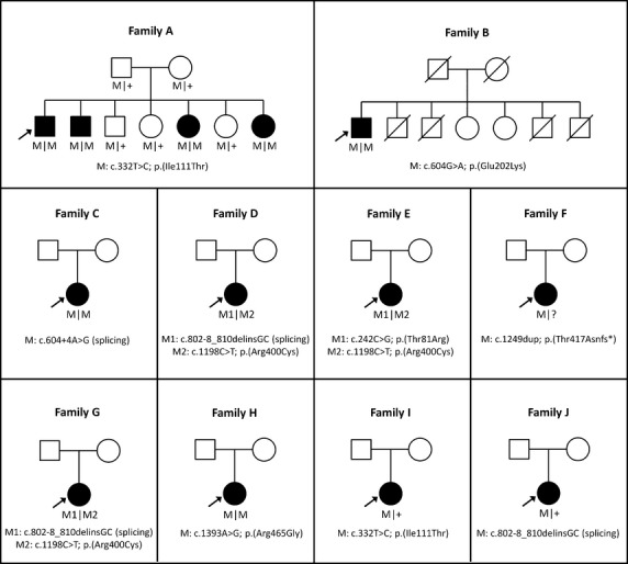
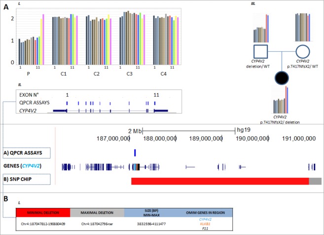
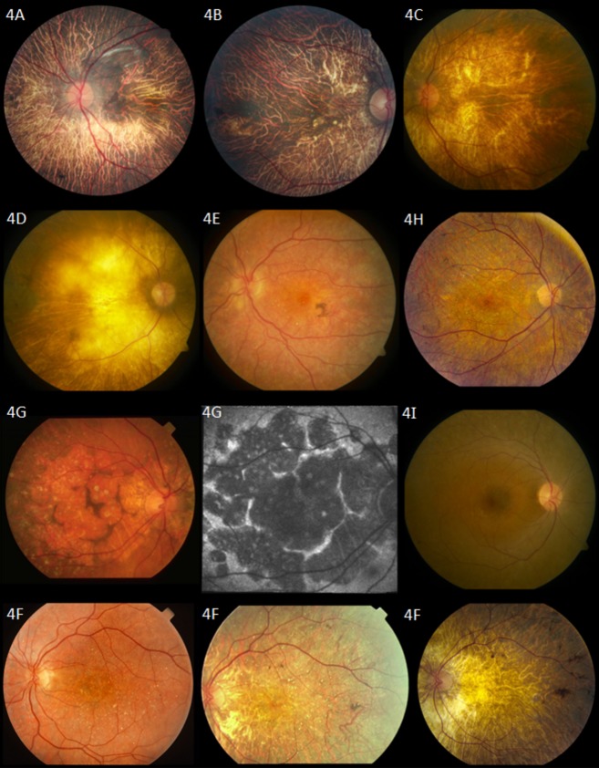
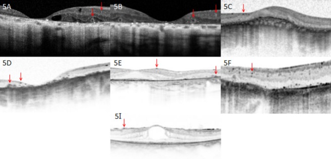
Bietti's crystalline dystrophy (BCD) is a rare, autosomal recessive retinal degenerative disease associated with mutations in CYP4V2. In this study, we describe the genetic and clinical findings in 19 unrelated BCD patients recruited from five international retinal dystrophy clinics. Patients underwent ophthalmic examinations and were screened for CYP4V2 mutations by Sanger sequencing and quantitative polymerase chain reaction (qPCR) copy number variation screening. Eight CYP4V2 mutations were found in 10/19 patients, including three patients in whom only monoallelic mutations were detected. Four novel mutations were identified: c.604G>A; p.(Glu202Lys), c.242C>G; p.(Thr81Arg), c.604+4A>G; p.(?), and c.1249dup; p.(Thr417Asnfs*2). In addition, we identified a heterozygous paternally inherited genomic deletion of at least 3.8 Mb, encompassing the complete CYP4V2 gene and several other genes, which is novel. Clinically, patients demonstrated phenotypic variability, predominantly showing choroidal sclerosis, attenuated vessels, and crystalline deposits of varying degrees of severity. To our knowledge, our study reports the first heterozygous CYP4V2 deletion and hence a novel mutational mechanism underlying BCD. Our results emphasize the importance of copy number screening in BCD. Finally, the identification of CYP4V2-negative patients with indistinguishable phenotypes from CYP4V2-positive patients might suggest the presence of mutations outside the coding regions of CYP4V2, or locus heterogeneity, which is unreported so far.

Keywords: Bietti; CYP4V2; crystalline dystrophy; retinal dystrophy.


Figures

Figure 1
Figure 1
Pedigrees of 10 families with Bietti crystalline dystropy carrying mutations in CYP4V2. Affected individuals are indicated with filled symbols, whereas unaffected relatives are indicated by open symbols. The probands are indicated with an arrow and slashed symbols indicate deceased. Mutated alleles are indicated with M symbols and wild-type allele with plus symbols.
Figure 2
Figure 2
Detection and delineation of a CYP4V2 deletion in proband F using qPCR and SNP-chip analysis. C (control), P (patient), WT (wild type). (A) I and II: qPCR on proband F revealed a copy number of one for all 11 assays of CYP4V2, corresponding with a heterozygous total gene deletion. The same was found for the other deleted genes; KLKB1 and F11. III: Demonstration of paternal origin of the deletion. (B) I: SNP-chip analysis demonstrates deletion with size ∼4 Mb, spanning the entire CYP4V2 gene and several other genes, two of which are OMIM genes (KLKB1 and F11).
Figure 3
Figure 3
Crystalline deposits seen on the lens (i) and corneal limbus (ii) of 54-year-old proband D with compound heterozygous mutations in CYP4V2 c.802-8_810delinsGC; p.(?), p.(Arg400Cys).
Figure 4
Figure 4
Fundus photographs. Proband A: 57-year-old patient with homozygous mutations in CYP4V2 p.(Ile111Thr). Fundus photograph shows choroidal sclerosis and peripheral pigmentation. No crystal deposits are seen in this patient's fundus. Proband B: 67-year-old patient with homozygous mutations in CYP4V2 p.(Glu202Lys). Fundus photograph shows severe choroidal sclerosis involving the entire macula and fovea, narrowing of the retinal vasculature, with relatively normal optic nerve appearance. Several crystalline deposits can be seen in the macula. Proband C: 47-year-old patient with homozygous mutation in CYP4V2 c.604+4A>G; p.(?). Severe choroidal sclerosis and crystalline deposits in macula. Proband D: 54-year-old patient with compound heterozygous mutations in CYP4V2 c.802-8_810delinsGC; p.(?), p.(Arg400Cys). Severe choroidal sclerosis and area of visible sclera. Few crystalline deposits seen in midperiphery. Proband E: A 41-year-old patient with compound heterozygous mutations in CYP4V2 p.(Thr81Arg), p.(Arg400Cys). Mild choroidal sclerosis, crystalline deposits, and pigment clump. Some maculopathy is seen. Retinal vasculature remains relatively normal. Proband F: 49-year-old patient with homozygous mutations in CYP4V2 p.(Thr417Nfs*2). Fundus photographs show progression of disease in left eye (left to right). Of note is the progressive choroidal sclerosis, vascular attenuation and decreasing presence of crystalline deposits over time. Photos taken at ages 31, 38, and 49, respectively. Proband G: 77-year-old patient with compound heterozygous mutation in CYP4V2 c.802-8_810delinsGC; p.(?), p.(Arg400Cys). Unusual fundus appearance (left) showing large, similarly-sized discoid patches of atrophy separated by small ridges of intact tissue with presence of crystals and choroidal sclerosis. Optic disk and retinal vasculature appear relatively normal. Fundus autofluorescence (middle) confirms areas of atrophy with patches of decreased autofluorescence surrounded by walls of lipofuscin metabolism. Proband H: 38-year-old patient with homozygous mutations in CYP4V2 p.(Arg465Gly). Choroidal sclerosis with diffuse crystalline deposits and pigment clumps in midperiphery. Proband I: 33-year-old patient with one heterozygous mutation in CYP4V2 p.(Ile111Thr). Fundus photograph reveals nondescript retina with few crystalline deposits.
Figure 5
Figure 5
Optical coherence tomography images. Proband A: 57-year-old patient with homozygous mutations in CYP4V2 p.(Ile111Thr). Optical coherence tomography shows severe foveal thinning and cystoid macular edema. Of note are two types of crystal that can be seen; tiny intraretinal crystals and large encapsulated subretinal crystals (red arrows). Proband B: 67-year-old patient with homozygous mutations in CYP4V2 p.(Glu202Lys). Shown are remarkable thinning of the fovea and remodeling of retina, as well as presence of both intraretinal and subretinal crystals. Proband C: 47-year-old patient with homozygous mutation in CYP4V2 c.604+4A>G; p.(?). Remodeling of retina with almost complete loss of architecture is seen, as well as tiny intraretinal crystals. Proband D: 54-year-old patient with compound heterozygous mutations in CYP4V2 c.802-8_810delinsGC; p.(?), p.(Arg400Cys). Optical coherence tomography shows severe foveal thinning, remodeling of the retina, as well as both intraretinal crystals and larger subretinal crystals. Proband E: A 41-year-old patient with compound heterozygous mutations in CYP4V2 p.(Thr81Arg), p.(Arg400Cys). Presence of both intraretinal and subretinal crystals. Photoreceptors in fovea remain intact. Proband F: 49-year-old patient with homozygous mutations in CYP4V2 p.(Thr417Nfs*2). Optical coherence tomography shows fine intraretinal crystals. Proband I: 33-year-old patient with one heterozygous mutation in CYP4V2 p.(Ile111Thr). Optical coherence tomography reveals presence of intraretinal crystals.

Similar articles

Cited by

References

    1. Bietti G. Ueber familiaeres vorkommen von “retinitis punctata albescens” (verbunden mit “dystrophia marginalis cristallinea corneae”), glitzern des glaskoerpers und anderen degenerativen augenveraenderungen. Klin. Monbl. Augenheilkd. 1937;99:21.
    1. Bolton-Maggs P, Wan-Yin B, McGraw A, Slack J. Kernoff P. Inheritance and bleeding in factor XI deficiency. Br. J. Haematol. 1988;69:521–528. - PubMed
    1. Chung DW, Fujikawa K, McMullen BA. Davie EW. Human plasma prekallikrein, a zymogen to a serine protease that contains four tandem repeats. Biochemistry. 1986;25:2410–2417. - PubMed
    1. Chung JK, Shin JH, Jeon BR, Ki CS. Park TK. Optical coherence tomographic findings of crystal deposits in the lens and cornea in Bietti crystalline corneoretinopathy associated with mutation in the CYP4V2 gene. Jpn. J. Ophthalmol. 2013;57:447–450. - PubMed
    1. Collin RW, van den Born LI, Klevering BJ, de Castro-Miró M, Littink KW, Arimadyo K, et al. High-resolution homozygosity mapping is a powerful tool to detect novel mutations causative of autosomal recessive RP in the Dutch population. Invest. Ophthalmol. 2011;52:2227–2239. - PubMed