. 2015 Jan;3(1):40-58.
doi: 10.1002/mgg3.111. Epub 2014 Sep 15.

Taurodontism, variations in tooth number, and misshapened crowns in Wnt10a null mice and human kindreds

Affiliations

Taurodontism, variations in tooth number, and misshapened crowns in Wnt10a null mice and human kindreds

Jie Yang et al. Mol Genet Genomic Med. 2015 Jan.

Abstract

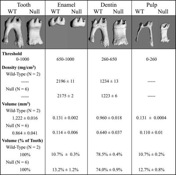
WNT10A is a signaling molecule involved in tooth development, and WNT10A defects are associated with tooth agenesis. We characterized Wnt10a null mice generated by the knockout mouse project (KOMP) and six families with WNT10A mutations, including a novel p.Arg104Cys defect, in the absence of EDA,EDAR, or EDARADD variations. Wnt10a null mice exhibited supernumerary mandibular fourth molars, and smaller molars with abnormal cusp patterning and root taurodontism. Wnt10a (-/-) incisors showed distinctive apical-lingual wedge-shaped defects. These findings spurred us to closely examine the dental phenotypes of our WNT10A families. WNT10A heterozygotes exhibited molar root taurodontism and mild tooth agenesis (with incomplete penetrance) in their permanent dentitions. Individuals with two defective WNT10A alleles showed severe tooth agenesis and had fewer cusps on their molars. The misshapened molar crowns and roots were consistent with the Wnt10a null phenotype and were not previously associated with WNT10A defects. The missing teeth contrasted with the presence of supplemental teeth in the Wnt10a null mice and demonstrated mammalian species differences in the roles of Wnt signaling in early tooth development. We conclude that molar crown and root dysmorphologies are caused by WNT10A defects and that the severity of the tooth agenesis correlates with the number of defective WNT10A alleles.

Keywords: Familial tooth agenesis; hypodontia; oligodontia, taurodontism.


Figures

Figure 1
Figure 1
Mandibular molar morphology in Wnt10a null mice at 16 weeks. (A) Radiographic images of the mandibular molars show significant root dysmorphology (taurodontism) in which the bifurcation of the roots is minimal or does not occur altogether. Rightward-pointing white arrowheads mark the level of root bifuration in the mandibular first molars. Failure of the roots to separate and extend mesially or distally precluded the development of interradicular alveolar bone, but expanded the interdental alveolar bone between the first and second molars (black arrowheads). Taurodontism is also evident on the mandibular second molars, whereas the third molars are normally single-rooted. (B) Dissecting microscope (top) and scanning electron microscope (bottom) images of the lateral aspects of mandibular molars. Fourth molars or residual sockets (downward-pointing white arrowheads) where the fourth molars had been lost were observed in all of the Wnt10a null mice. (C) Dissecting microscope (top) and scanning electron microscope (bottom) images of the occlusal aspects of the mandibular molars. The null first molars were characterized by smaller, more rounded crowns lacking a distal cusp and by the presence of a prominent central cusp delineated by a deep groove (upward-pointing arrowheads). In the case of the 1316 null mice, the central groove was stained and apparently carious, something we have never before observed in laboratory mice. The molar occlusal surfaces of the Wnt10a null mice did not show any signs of attrition that would be expected if the functional properties of enamel or dentin had been reduced as a consequence of developing in the absence of WNT10A.
Figure 2
Figure 2
SEMs of extracted mandibular first molars at 16 weeks. Altered crown and root morphology are apparent in images taken at 30×. Higher magnitude (200×) images show the texture of the root surface below the dentino-enamel junction (DEJ, arrowheads). The 600× images magnify the boxed regions from the 200× views. Resorption lacunae of irregular in size and shape were present on the roots of the Wnt10a null molars, but not on the wild-type molar.
Figure 3
Figure 3
MicroCT images of the right and left mandibular first molars from 16-week-old wild-type and Wnt10a null mice. The 3D μCT images were generated for the mandibular first molars scanned in situ (within the hemimandibles). The teeth were outlined to focus on the dental structures. Hard tissue and soft tissues were imaged by setting the threshold above or below 260, respectively. The sagittal views revealed irregular mineral structures that corresponded to vacancies within pulpal tissue (arrowheads). These are pulp calcifications or “pulp stones”, which are more extensive in the molars of the Wnt10a null mice than in the wild type. Raising the threshold above 260 did not selectively remove the pulp stones, indicating that they are approximately the same density as dentin.
Figure 4
Figure 4
MicroCT analyses of mineral density and volume of mandibular first molars at 16 weeks. The whole tooth, enamel, dentin, and pulp were individually isolated by varying the density threshold for detection. The density measurements were similar in the Wnt10a null and wild-type mice, with the density of enamel and dentin in the null molars being over 99% as dense as the enamel and dentin of the wild type. The null first molars were smaller than the wild type: having only 71% as much volume. The reduction in the volume of dentin was slightly greater than the reductions in the volumes of enamel and pulp, so that the enamel and pulp comprised about 2.5% and 2% more of the volume of null molars compared to that of the wild type, respectively.
Figure 5
Figure 5
MicroCT analysis of mandibular incisors at 16 weeks. (A) 3D reconstructions of mandibular incisors at 16 weeks. Orientations: The apical ends of the incisors are toward the center; incisal tips are toward the periphery; lingual (dentin) side is up (toward the viewer); buccal (enamel) side is down and not visible. All incisors from the Wnt10a null mice showed a failure to close in their apical–lingual region. In Null 1316 a single wedge-shaped defect extended from the apical end and narrowed incisally. In the Null 1317 and Null 1329 mice, the defects extended further toward the middle of the incisors, with a thin strip of dentin running at the center of the defects. Thus, in the Null 1316, there is a single wedge-shaped deficiency, whereas in Null 1317 and Null 1329, there are two overlapping wedge-shaped deficiencies. White arrowheads and black arrowheads mark the positions of 2D cross sections shown in the top and bottom rows of part B, respectively. (B) Cross sections from apical ends of the incisors. The wild-type incisor has a closed circle of dentin at its apical end. The Wnt10a Null 1316 incisor cross sections shows a single gap on the lingual aspect of the mandibular incisor that narrows from apical to incisal. The Null 1317 and 1329 incisor cross sections show two openings on the lingual face corresponding to the two wedge-shaped deficiencies in the lingual dentin.
Figure 6
Figure 6
Pedigrees and anomalous dental morphologies in persons with WNT10A defects: Families 1 and 2. (A) Pedigree of Family 1. A dot marks each of the eight individuals that were recruited and evaluated. The WNT10A genotype is shown for each recruited member. Only the proband (III:6) had developmentally absent teeth (18 permanent teeth) and was the only individual with defects in both WNT10A alleles (p.Arg104Cys/p.Gly213Ser). (B) Detail from the proband's (III:6) panorex radiograph showing taurodontism in all first molars (#3, 14, 19, 30) (top). Oral photograph showing the proband's misshapened central incisors (bottom). (C) Oral photograph of proband's maternal grandfather (I:3), who was heterozygous for the p.R104C defect in WNT10A, showing peg lateral incisors (#7, 10). (D) Details from the panorex radiograph of the proband's younger sister (III:7), who was heterozygous for the p.Arg104Cys defect in WNT10A, showing taurodontism in all primary mandibular first molars (#K, L, S, T). (E) Pedigree of Family 2. A dot marks each of the six individuals that were recruited and evaluated. The WNT10A genotype is shown for each recruited member. Affected members had developmentally missing teeth: III:2 (2 teeth); III:3 (1 tooth); IV:4 (8 teeth); IV:5 (13 teeth). (F) Details from the panorex radiograph of the proband's maternal aunt (III:2), who was heterozygous for the p.Cys107*WNT10A defect, showing taurodontism in the mandibular second molars (#17, 31). (G) Details from the panorex radiograph of the proband's mother (III:3), who was heterozygous for the p.Cys107*WNT10A defect, showing taurodontism in the mandibular second molars (#18, 31). (H) Details from the proband's (IV:4) panorex radiograph, who had defects in both WNT10A alleles (p.Cys104*/p.Phe228Ile), showing peg lateral incisors (left; #7, 10) and taurodontism in the mandibular first molars (right; #19, 30). Oral photographs show rounded first molar morphology, with maxillary first molars (left; #3, 15) lacking the distal palatal cusp and the mandibular first molars (right; #19, 30) lacking the distal cusp. (I) Details from the proband's younger sister's (IV:5) panorex radiograph, who also had defects in both WNT10A alleles (p.Cys104*/p.Phe228Ile), showing taurodontism in the primary mandibular first molars (right; #L, S). Oral photographs show rounded first molar morphology, with maxillary first molars (left; #3, 15) lacking the distal palatal cusp and the mandibular first molars (right; 19, 30) lacking the distal cusp.
Figure 7
Figure 7
Pedigrees and anomalous dental morphologies in persons with WNT10A defects: Families 3 through 6. (A) Pedigree of Family 3. A dot marks each of the five individuals that were recruited and evaluated. The WNT10A genotype is shown for each recruited member. All four WNT10A alleles in the proband's parents carried different WNT10A sequence variations. Affected members with developmentally missing teeth were: II:3 (6 teeth); II:4 (2 teeth); III:1 (23 teeth). (B) Details from the panorex radiograph of the proband's father (II:3) showing taurodontism in the mandibular second molars (#18, 31). (C) Details from the proband's (III:2) panorex radiograph showing conical maxillary central incisors (top; #8, 9) taurodontism in all primary first and second molars (#A, B, I, J, K, L, S, T). (D) Details from the panorex radiograph of the proband's younger brother (III:2) showing taurodontism in the mandibular first molars (#19, 30). (E) Pedigree of Family 4 and oral photograph of the proband (II:1) who had 17 developmentally absent teeth and peg-shaped maxillary incisors (#7, 8, 9, 10). (F) Pedigree of Family 5 and detail from the proband's (II:1) panorex radiograph showing taurodontism of the mandibular second molars (#18, 31). The proband had 11 developmentally absent teeth and peg-shaped maxillary central incisors (#8, 9). (G) Pedigree of Family 6 and detail from the proband's (II:1) panorex radiograph showing taurodontism of the mandibular first molars (#19, 30). The proband had three developmentally absent teeth.

Similar articles

Cited by

References

    1. Adaimy L, Chouery E, Megarbane H, Mroueh S, Delague V, Nicolas E, et al. Mutation in WNT10A is associated with an autosomal recessive ectodermal dysplasia: the odonto-onycho-dermal dysplasia. Am. J. Hum. Genet. 2007;81:821–828. - PMC - PubMed
    1. Arte S, Parmanen S, Pirinen S, Alaluusua S. Nieminen P. Candidate gene analysis of tooth agenesis identifies novel mutations in six genes and suggests significant role for WNT and EDA signaling and allele combinations. PLoS ONE. 2013;8:e73705. - PMC - PubMed
    1. Bae CH, Lee JY, Kim TH, Baek JA, Lee JC, Yang X, et al. Excessive Wnt/beta-catenin signaling disturbs tooth-root formation. J. Periodontal Res. 2013;48:405–410. - PubMed
    1. Bohring A, Stamm T, Spaich C, Haase C, Spree K, Hehr U, et al. WNT10A mutations are a frequent cause of a broad spectrum of ectodermal dysplasias with sex-biased manifestation pattern in heterozygotes. Am. J. Hum. Genet. 2009;85:97–105. - PMC - PubMed
    1. van den Boogaard MJ, Creton M, Bronkhorst Y, van der Hout A, Hennekam E, Lindhout D, et al. Mutations in WNT10A are present in more than half of isolated hypodontia cases. J. Med. Genet. 2012;49:327–331. - PubMed