Review
. 2015:2015:808314.
doi: 10.1155/2015/808314. Epub 2015 Jan 5.

Management of cerebral cavernous malformations: from diagnosis to treatment

Affiliations
Review

Management of cerebral cavernous malformations: from diagnosis to treatment

Nikolaos Mouchtouris et al. ScientificWorldJournal. 2015.

Abstract

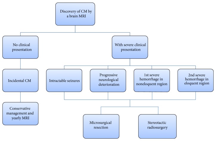
Cerebral cavernous malformations are the most common vascular malformations and can be found in many locations in the brain. If left untreated, cavernomas may lead to intracerebral hemorrhage, seizures, focal neurological deficits, or headaches. As they are angiographically occult, their diagnosis relies on various MR imaging techniques, which detect different characteristics of the lesions as well as aiding in planning the surgical treatment. The clinical presentation and the location of the lesion are the most important factors involved in determining the optimal course of treatment of cavernomas. We concisely review the literature and discuss the advantages and limitations of each of the three available methods of treatment--microsurgical resection, stereotactic radiosurgery, and conservative management--depending on the lesion characteristics.


Figures

Figure 1
Figure 1
Flow-chart of the work-up and management of patients with cavernous malformations. Once a cavernomas is diagnosed via an MRI of the brain, deciding the course of treatment depends on the clinical presentation of the patient. Purely incidental cavernomas are managed conservatively and followed by yearly MRI scans. Cavernomas are treated by microsurgical resection or stereotactic radiosurgery if the patient is experiencing severe symptoms, such as intractable seizures, progressive neurological deterioration, one severe hemorrhage in a noneloquent region of the brain, or at least two severe hemorrhages in eloquent brain. Selecting between resection and radiosurgery depends on the location of the lesion and the severity of the presentation as explained in this paper.

References

    1. Dalyai R. T., Ghobrial G., Awad I., et al. Management of incidental cavernous malformations: a review. Neurosurgical Focus. 2011;31(6, article E5) doi: 10.3171/2011.9.FOCUS11211. - DOI - PubMed
    1. Yadla S., Jabbour P. M., Shenkar R., Shi C., Campbell P. G., Awad I. A. Cerebral cavernous malformations as a disease of vascular permeability: from bench to bedside with caution. Neurosurgical focus. 2010;29(3):p. E4. doi: 10.3171/2010.5.FOCUS10121. - DOI - PMC - PubMed
    1. Raychaudhuri R., Batjer H. H., Awad I. A. Intracranial cavernous angioma: a practical review of clinical and biological aspects. Surgical Neurology. 2005;63(4):319–328. doi: 10.1016/j.surneu.2004.05.032. - DOI - PubMed
    1. D'Angelo V. A., De Bonis C., Amoroso R., et al. Supratentorial cerebral cavernous malformations: clinical, surgical, and genetic involvement. Neurosurgical Focus. 2006;21(1):7. - PubMed
    1. Wang C.-C., Liu A., Zhang J.-T., Sun B., Zhao Y.-L. Surgical management of brain-stem cavernous malformations: report of 137 cases. Surgical Neurology. 2003;59(6):444–454. doi: 10.1016/S0090-3019(03)00187-3. - DOI - PubMed

MeSH terms

LinkOut - more resources