. 2015 Jan 28;10(1):e0114932.
doi: 10.1371/journal.pone.0114932. eCollection 2015.

Rapid screening of gene function by systemic delivery of morpholino oligonucleotides to live mouse embryos

Affiliations

Rapid screening of gene function by systemic delivery of morpholino oligonucleotides to live mouse embryos

Kathryn S McClelland et al. PLoS One. .

Abstract

Traditional gene targeting methods in mice are complex and time consuming, especially when conditional deletion methods are required. Here, we describe a novel technique for assessing gene function by injection of modified antisense morpholino oligonucleotides (MOs) into the heart of mid-gestation mouse embryos. After allowing MOs to circulate through the embryonic vasculature, target tissues were explanted, cultured and analysed for expression of key markers. We established proof-of-principle by partially phenocopying known gene knockout phenotypes in the fetal gonads (Stra8, Sox9) and pancreas (Sox9). We also generated a novel double knockdown of Gli1 and Gli2, revealing defects in Leydig cell differentiation in the fetal testis. Finally, we gained insight into the roles of Adamts19 and Ctrb1, genes of unknown function in sex determination and gonadal development. These studies reveal the utility of this method as a means of first-pass analysis of gene function during organogenesis before committing to detailed genetic analysis.


Conflict of interest statement

Competing Interests: The authors have declared that no competing interests exist.

Figures

Fig 1
Fig 1. Overview of method: MO delivery by heart injection.
(A) Experimental pipeline from harvest of embryos through to injection, culture and downstream analyses. Visualisation of heart injection protocol can be seen in S1 Video and images B–E. The cocktail of dye and MO in PBS is delivered via injection into the left ventricle of the beating heart at 11.5 dpc (B). Dye can be visualised going around the embryonic vasculature (indicated by white arrows) (C, D) and into the head vasculature (D) before the whole embryo is coloured (E). Schematic of ventricle injection (F) and the embryonic gonad which is highly vascularised (G). Delivery of India ink and F-MO (indicated by white arrows) shows the compounds reaching the mesonephric plexus at 5 min post-injection (H; n = 3); after 30 min F-MO positive cells were observed in the gonad proper (I; n = 3). s = seconds; min = minutes; g = gonad; m = mesonephros; F-MO = carboxyfluorescein-labelled standard control morpholino oligonucleotide. Scale bars: E = 1 mm, H = 0.5 mm.
Fig 2
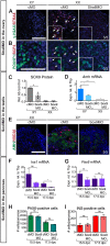
Fig 2. Partial phenocopy of known gene knockouts in gonad and pancreas.
(A, B) STRA8 knockdown: IF showed knockdown of STRA8 (A) in Stra8MO-treated XX gonads. Nuclear localisation of meiosis markers (γH2AX (A) and SCP3 (B); indicated by white arrows; see inserts) was absent but germ cells were present (POU5F1 (B); see inserts) in XX Stra8MO-treated gonads. (C–E) Knockdown of SOX9 in the gonad: Western blot for SOX9 (relative to α-TUBULIN or β-ACTIN) showed a downregulation of SOX9 (C) after Sox9MO treatment in XY gonads (n = 3). Downregulation of expression of SOX9 target gene Amh (D) expression was observed by qRT-PCR (n = 8, 15, 11, 4). IF for AMH and HSD3β (E) showed that AMH staining was weaker in XY Sox9MO samples compared to XY controls and that HSD3β-positive FLCs were present but staining was weaker in XY Sox9MO-treated gonads. (F–I) Knockdown of SOX9 in the pancreas: qRT-PCR of Sox9Mo treated pancreata showed Ins1 (F) was downregulated but Pax6 (G) was unchanged (n = 5, 5, 5, 5). Quantification of PAX6/INS-positive cells revealed that PAX6-positive (H) and INS-positive (I) cell number was unaltered by Sox9MO treatment (n = 3, 4, 2, 2). Scale bars = 100 μM; cMO = control morpholino; xMO = morpholino targeting gene x. For Western blots SOX9 levels were normalised to α-TUBULIN or β-ACTIN loading controls and Sox9MO-treated XY gonads measured relative to cMO treated XY gonads with expression for each blot set to 1. Rel. Ab./control = Relative Abundance of SOX9 to α-TUBULIN or β-ACTIN. For all qRT-PCR levels are shown relative to Tbp, error = S.E.M. For cell quantification error = S.E.M. with individual counts plotted. * = p = 0.05, ** = p = 0.001, *** = p = 0.0001, ns = not statistically significant.
Fig 3
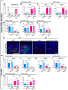
Fig 3. Double knockdown of Gli1/Gli2 in XY gonads.
(A–D) Knockdown of GLI1/GLI2 in the gonad: qRT-PCR showed that treatment with Gli1/Gli2MO (n = 6, 5, 5, 8) resulted in no significant downregulation in steroidogenic regulator Sf1/Nr5a1 (A) but a significant downregulation in expression of steroidogenic pathway enzymes Hsd3β (B), Cyp11a1 (C) and Star (D). No change was observed in Nr5a1 expression in Gli1MO or Gli2MO knockdown (E, I). Similarly, there were no changes in expression of steroidogenic pathway enzymes Hsd3β (F, J), Cyp11a1 (G, K) and Star (H, L) in Gli1MO (E–H; n = 6, 6, 7, 5) or Gli2MO (I–L; n = 8, 7, 4, 3) single knockdowns. IF showed Sertoli cells (AMH (M) and SOX9 (N)) and germ cells (POU5F1 (M)) were present in XY Gli1/Gli2MO treated gonads and no FOXL2-positive cells were observed (N). Steroidogenic Hsd3β-positive (M) and Nr5a1-positive (N) cells were still present in Gli1/Gli2MO treated XY gonads. Quantification (n = 2) of steroidogenic cells revealed no change in the number of HSD3β-positive Leydig cells (O; green) or SF1-positive/SOX9-negative pre-Leydig cells (O; red). There was a decrease in the number of SOX9-positve Sertoli cells in the Gli1/2MO treated XY gonads (O; yellow). Scale bars = 100 μM; cMO = control morpholino; xMO = morpholino targeting gene x. For all qRT-PCR levels are shown relative to Tbp, error = S.E.M. For cell quantification error = S.E.M. with individual counts plotted. * = p = 0.05, ** = p = 0.001, ns = not statistically significant.
Fig 4
Fig 4. Knockdown of Adamts19 in XX gonads and Ctrb1 in XY gonads.
(A) qRT-PCR on FACS-sorted somatic and germ cells (n = 3, 4, 3, 4) shows that Adamts19 is expressed in the somatic cells of the ovary at 12.5 dpc and at much lower levels in somatic cells of the testis. Knockdown of ADAMTS19 in the XX gonad (n = 7, 8, 5, 5) showed no change in female somatic markers Fst (B) and Irx3 (C) and a slight decrease in expression of germ cell marker Ddx4 (D). Male markers, Amh (Sertoli cells; E), Nr5a1 (Somatic cells; F) and Cyp11a1 (Leydig cells; G) were unperturbed. IF showed no discernable difference in the ratios of FOXL2-positive/DDX4-positive cells in the Adamts19MO-treated XX gonad compared to the control (H). Knockdown of CTRB1 in the XY gonad (n = 19, 16, 14, 14) resulted in no change to male somatic markers Sox9 (I) or Amh (J) but an increase in Ptgds (K) was observed in the Ctrb1MO-treated XY gonad. Expression of Leydig cell marker Cyp11a1 (L), female somatic marker Fst (M) and germ cell marker Ddx4 (N) was unchanged. Germ = germ cells, Som. = somatic cells. Scale bars = 100 μM; cMO = control morpholino; xMO = morpholino targeting gene x. For all qRT-PCR: levels are shown relative to Tbp, error = S.E.M., * = p = 0.05, ** = p = 0.001, *** = p = 0.0001, **** = p = 0.00001, ns = not statistically significant.

References

    1. Hsu PD, Lander ES, Zhang F (2014) Development and Applications of CRISPR-Cas9 for Genome Engineering. Cell 157: 1262–1278. 10.1016/j.cell.2014.05.010 - DOI - PMC - PubMed
    1. Nakamura Y, Yamamoto M, Matsui Y (2002) Introduction and expression of foreign genes in cultured mouse embryonic gonads by electroporation. Reprod Fertil Dev 14: 259–265. 10.1071/RD01130 - DOI - PubMed
    1. Ryan J, Ludbrook L, Wilhelm D, Sinclair A, Koopman P, et al. (2011) Analysis of gene function in cultured embryonic mouse gonads using nucleofection. Sex Dev 5: 7–15. 10.1159/000322162 - DOI - PubMed
    1. Svingen T, Wilhelm D, Combes AN, Hosking B, Harley VR, et al. (2009) Ex vivo magnetofection: A novel strategy for the study of gene function in mouse organogenesis. Dev Dyn 238: 956–964. 10.1002/dvdy.21919 - DOI - PMC - PubMed
    1. Dean CH, Miller L-AD, Smith AN, Dufort D, Lang RA, et al. (2005) Canonical Wnt signaling negatively regulates branching morphogenesis of the lung and lacrimal gland. Dev Biol 286: 270–286. 10.1016/j.ydbio.2005.07.034 - DOI - PubMed

Publication types

MeSH terms