. 2015 Jan 28;10(1):e0117908.
doi: 10.1371/journal.pone.0117908. eCollection 2015.

TGFβ signaling in myeloid cells regulates mammary carcinoma cell invasion through fibroblast interactions

Affiliations

TGFβ signaling in myeloid cells regulates mammary carcinoma cell invasion through fibroblast interactions

Aubie K Shaw et al. PLoS One. .

Abstract

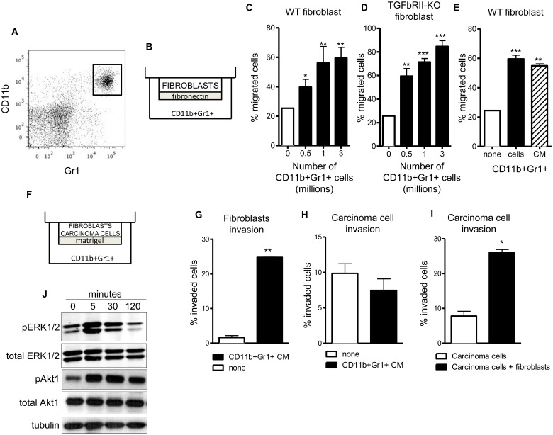
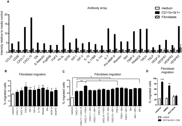
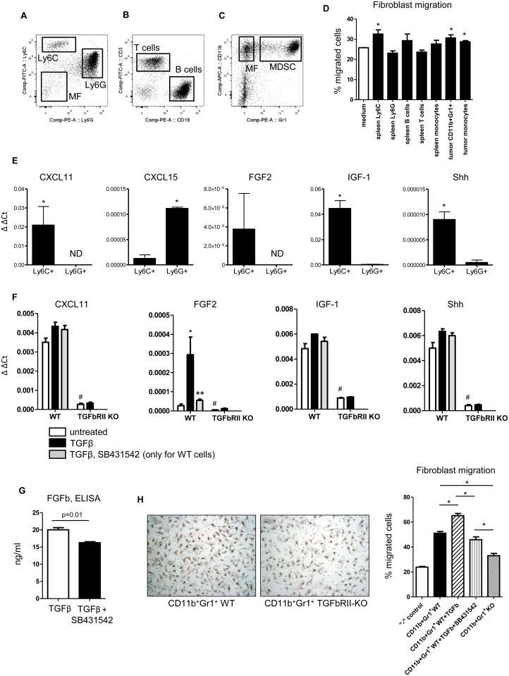
Metastasis is the most devastating aspect of cancer, however we know very little about the mechanisms of local invasion, the earliest step of metastasis. During tumor growth CD11b+ Gr1+ cells, known also as MDSCs, have been shown to promote tumor progression by a wide spectrum of effects that suppress the anti-tumor immune response. In addition to immunosuppression, CD11b+ Gr1+ cells promote metastasis by mechanisms that are currently unknown. CD11b+ Gr1+ cells localize near fibroblasts, which remodel the ECM and leave tracks for collective cell migration of carcinoma cells. In this study we discovered that CD11b+ Gr1+ cells promote invasion of mammary carcinoma cells by increasing fibroblast migration. This effect was directed by secreted factors derived from CD11b+ Gr1+ cells. We have identified several CD11b+ Gr1+ cell secreted proteins that activate fibroblast migration, including CXCL11, CXCL15, FGF2, IGF-I, IL1Ra, Resistin, and Shh. The combination of CXCL11 and FGF2 had the strongest effect on fibroblast migration that is associated with Akt1 and ERK1/2 phosphorylation. Analysis of subsets of CD11b+ Gr1+ cells identified that CD11b+ Ly6Chigh Ly6Glow cells increase fibroblast migration more than other myeloid cell populations. Additionally, tumor-derived CD11b+ Gr1+ cells promote fibroblast migration more than splenic CD11b+ Gr1+ cells of tumor-bearing mice. While TGFβ signaling in fibroblasts does not regulate their migration toward CD11b+ Gr1+ cells, however deletion of TGFβ receptor II on CD11b+ Gr1+ cells downregulates CXCL11, Shh, IGF1 and FGF2 resulting in reduced fibroblast migration. These studies show that TGFβ signaling in CD11b+ Gr1+ cells promotes fibroblast directed carcinoma invasion and suggests that perivascular CD11b+ Ly6Chigh Ly6Glow cells may be the stimulus for localized invasion leading to metastasis.


Conflict of interest statement

Competing Interests: The authors have declared that no competing interests exist.

Figures

Fig 1
Fig 1. CD11b+Gr1+ cells increase migration of fibroblasts and invasion of co-cultured carcinoma cells.
(A) CD11b+Gr1+ cells were isolated from spleen of tumor-bearing mice by magnetic separation and cultured for 16–18 hours. (B) Fibronectin-coated 8 uM pore Transwells were placed on top of cultured CD11b+Gr1+ cells and 5x10⁴ mammary fibroblasts were allowed to migrate for 5 hours. (C) CD11b+Gr1+ cells dose-dependently increase fibroblast migration. (D) Fibroblasts with deletion of TGFβRII have a further increase in migration in response to CD11b+Gr1+ cells. (E) Conditioned medium (CM) collected from CD11b+Gr1+ cells increases fibroblast migration to the same extent as live CD11b+Gr1+ cells. * – p<0.05, ** – p<0.01, *** – p<0.001 compared to no CD11b+Gr1+ cells. (F) Matrigel-coated 8 uM pore Transwells were placed on top of CD11b+Gr1+ CM and mammary carcinoma cells, fibroblasts or both cell types were allowed to invade for 18 hours. (G) CD11b+Gr1+ cells increase fibroblasts invasion. (H) CD11b+Gr1+ cells do not increase the invasion of carcinoma cells. (I) CD11b+Gr1+ cells increase carcinoma cell invasion when fibroblasts are co-cultured. * – p<0.05, ** – p<0.01, compared to open bars. (J) Western immunoblot of fibroblast Erk1/2 and Akt phosphorylation shows activation after 5 minutes of treatment with CM of CD11b+Gr1+ cells.
Fig 2
Fig 2. Cytokines and chemokines secreted by CD11b+Gr1+ cells increase fibroblast migration.
(A) Antibody array analysis of proteins secreted by CD11b+Gr1+ cells compared to fibroblasts. Although 144 proteins were assayed, only proteins predominantly expressed in CD11b+Gr1+ cells are shown. (B) Purified cytokine treatment of fibroblasts increases migration through Transwell chambers. (C) Combinations of several cytokines and chemokines increase fibroblast migration beyond treatment with a single cytokine/chemokine. Concentrations are same as on (B), * – p<0.05, ** – p<0.01, *** – p<0.001 compared to untreated; #: p<0.01 compared to all other treatments. (D) Inhibition of FGFR1 Kinase by PD173074 (5 nM) prevents fibroblast migration towards conditioned medium from CD11b+Gr1+ cells. ** – p<0.01, *** – p<0.001 relative to control medium.
Fig 3
Fig 3. TGFβRII signaling in Ly6ChighLy6Glow cells regulates the secretion of chemokines and results in increased fibroblast migration.
(A) FACS plot showing collection of Ly6C+, Ly6G+ and monocytes from spleen of 4T1 tumor-bearing mice. The plot was gated for CD45+ and CD11b+ cells. (B) FACS plot showing collection of T cells (CD3+) and B cells (CD19+) from spleen of 4T1 tumor-bearing mice. The plot was gated for CD45+ cells. (C) FACS plot showing collection of CD11b+Gr1+ cells and macrophages (CD11b+Gr1+) from 4T1 tumor tissue. The plot was gated for CD45+ cells. (D) Fibroblast migration to conditioned medium (from 1x10⁶ cells) prepared from subsets of splenocytes or myeloid cells from spleen and tumor tissue. Ly6C+ cells from spleen, tumor-derived CD11b+Gr1+ cells and tumor macrophages induce the most fibroblast migration. * – p<0.05 compared to control. (E) qRT-PCR analysis of cytokines/chemokines in splenic Ly6G (CD11b+Ly6GhighLy6Clow) and Ly6C (CD11b+Ly6ChighLy6Glow) cells. (F) qRT-PCR analysis of cytokines/chemokines in splenic CD11b+Gr1+ cells stimulated by TGFβ1 (1 ng/ml) for 18hr. with and without SB431542 (10 uM) * – p<0.05 compared with untreated cells, ** – p<0.05 compared with TGFβ treated cells, # – p<0.05 compared with WT cells. (G) CD11b+Gr1+ cells (4x10⁶) isolated from spleen of LLC tumor bearing mice were incubated 18hr in presence of TGFβ (1ng/ml) and TGFβ and SB431542 (10uM). Level of FGFb was measured by ELISA (R&D System, Minneapolis, MN). (H) CD11b+Gr1+ cells (3x10⁶) isolated from spleen of LLC tumor bearing mice with deleted TGFβRII decrease migration of fibroblasts compared with CD11b+Gr1+ cells with intact TGFβ signaling. “-” control—negative control, DMEM without serum, * – p<0.01. TGFβ1 (1 ng/ml), SB431542 (10 uM).

Similar articles

Cited by

References

    1. Coghlin C, Murray GI (2010) Current and emerging concepts in tumour metastasis. The Journal of pathology 222: 1–15. 10.1002/path.2727 - DOI - PubMed
    1. Brewster AM, Hortobagyi GN, Broglio KR, Kau SW, Santa-Maria CA, et al. (2008) Residual risk of breast cancer recurrence 5 years after adjuvant therapy. Journal of the National Cancer Institute 100: 1179–1183. 10.1093/jnci/djn233 - DOI - PMC - PubMed
    1. Kedrin D, Gligorijevic B, Wyckoff J, Verkhusha VV, Condeelis J, et al. (2008) Intravital imaging of metastatic behavior through a mammary imaging window. Nature methods 5: 1019–1021. 10.1038/nmeth.1269 - DOI - PMC - PubMed
    1. Mognetti B, La Montagna G, Perrelli MG, Pagliaro P, Penna C (2013) Bone marrow mesenchymal stem cells increase motility of prostate cancer cells via production of stromal cell-derived factor-1alpha. Journal of cellular and molecular medicine 17: 287–292. 10.1111/jcmm.12010 - DOI - PMC - PubMed
    1. Richardsen E, Sorbye SW, Crowe JP, Yang JL, Busund LT (2009) Expression of M-CSF and CSF-1R is correlated with histological grade in soft tissue tumors. Anticancer research 29: 3861–3866. - PubMed

Publication types

MeSH terms

Substances