

Novel localization of peripherin 2, the photoreceptor-specific retinal degeneration slow protein, in retinal pigment epithelium
- PMID: 25629227
- PMCID: PMC4346858
- DOI: 10.3390/ijms16022678
Novel localization of peripherin 2, the photoreceptor-specific retinal degeneration slow protein, in retinal pigment epithelium
Abstract
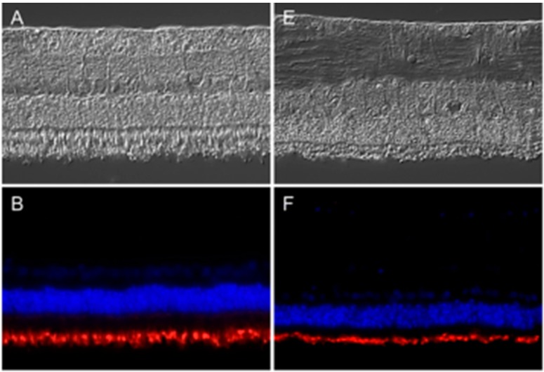
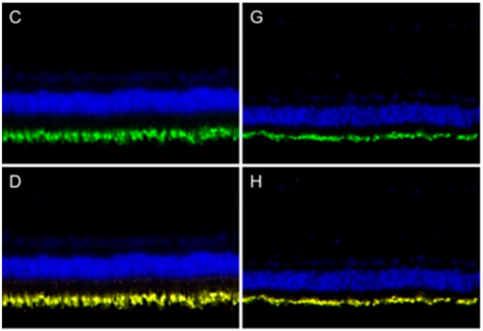
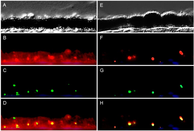
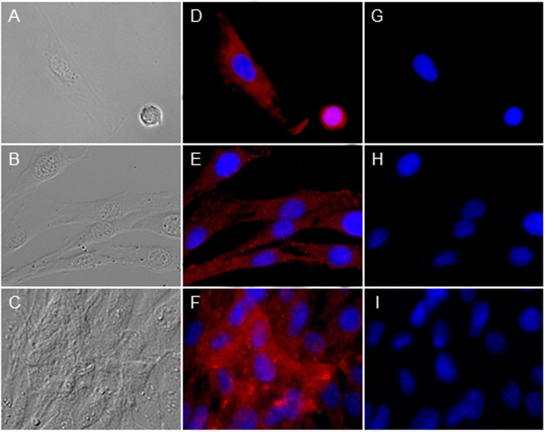
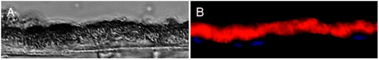
Retinal pigment epithelium (RPE) builds the outer blood-retinal barrier of the eye. Since one typical feature of the autoimmune disease, equine recurrent uveitis (ERU), is the breakdown of this barrier, we recently performed comparative analysis of healthy and uveitic RPE. We identified for the first time peripherin 2, which is responsible for visual perception and retina development, to be localized in RPE. The purpose of this study was therefore to validate our findings by characterizing the expression patterns of peripherin 2 in RPE and retina. We also investigated whether peripherin 2 expression changes in ERU and if it is expressed by the RPE itself. Via immunohistochemistry, significant downregulation of peripherin 2 in uveitic RPE compared to the control was detectable, but there was no difference in healthy and uveitic retina. A further interesting finding was the clear distinction between peripherin 2 and the phagocytosis marker, rhodopsin, in healthy RPE. In conclusion, changes in the expression pattern of peripherin 2 selectively affect RPE, but not retina, in ERU. Moreover, peripherin 2 is clearly detectable in healthy RPE due to both phagocytosis and the expression by the RPE cells themselves. Our novel findings are very promising for better understanding the molecular mechanisms taking place on RPE in uveitis.
Figures
Similar articles
-
In situ cell surface proteomics reveals differentially expressed membrane proteins in retinal pigment epithelial cells during autoimmune uveitis.J Proteomics. 2014 Sep 23;109:50-62. doi: 10.1016/j.jprot.2014.06.020. Epub 2014 Jul 2. J Proteomics. 2014. PMID: 24998980
-
CaV1.3 L-type channels, maxiK Ca(2+)-dependent K(+) channels and bestrophin-1 regulate rhythmic photoreceptor outer segment phagocytosis by retinal pigment epithelial cells.Cell Signal. 2014 May;26(5):968-78. doi: 10.1016/j.cellsig.2013.12.021. Epub 2014 Jan 7. Cell Signal. 2014. PMID: 24407175
-
miR-184 regulates ezrin, LAMP-1 expression, affects phagocytosis in human retinal pigment epithelium and is downregulated in age-related macular degeneration.FEBS J. 2014 Dec;281(23):5251-64. doi: 10.1111/febs.13066. Epub 2014 Oct 13. FEBS J. 2014. PMID: 25251993
-
Barrier properties of cultured retinal pigment epithelium.Exp Eye Res. 2014 Sep;126:16-26. doi: 10.1016/j.exer.2013.12.018. Epub 2014 Apr 14. Exp Eye Res. 2014. PMID: 24731966 Review.
-
Equine recurrent uveitis--a spontaneous horse model of uveitis.Ophthalmic Res. 2008;40(3-4):151-3. doi: 10.1159/000119867. Epub 2008 Apr 18. Ophthalmic Res. 2008. PMID: 18421230 Review.
Cited by
-
Immunological Insights in Equine Recurrent Uveitis.Front Immunol. 2021 Jan 8;11:609855. doi: 10.3389/fimmu.2020.609855. eCollection 2020. Front Immunol. 2021. PMID: 33488614 Free PMC article. Review.
-
High-Salt Enhances the Inflammatory Response by Retina Pigment Epithelium Cells following Lipopolysaccharide Stimulation.Mediators Inflamm. 2015;2015:197521. doi: 10.1155/2015/197521. Epub 2015 Dec 10. Mediators Inflamm. 2015. PMID: 26783382 Free PMC article.
References
-
- Uhl P.B., Szober C.M., Amann B., Alge-Priglinger C., Ueffing M., Hauck S.M., Deeg C.A. In situ cell surface proteomics reveals differentially expressed membrane proteins in retinal pigment epithelial cells during autoimmune uveitis. J. Proteomics. 2014;109:50–62. doi: 10.1016/j.jprot.2014.06.020. - DOI - PubMed
Publication types
MeSH terms
Substances
LinkOut - more resources
Full Text Sources
Other Literature Sources
