. 2015 Mar 15;191(6):626-36.
doi: 10.1164/rccm.201408-1565OC.

Wogonin induces eosinophil apoptosis and attenuates allergic airway inflammation

Affiliations

Wogonin induces eosinophil apoptosis and attenuates allergic airway inflammation

Christopher D Lucas et al. Am J Respir Crit Care Med. .

Abstract

Rationale: Eosinophils are key effector cells in allergic diseases, including allergic rhinitis, eczema, and asthma. Their tissue presence is regulated by both recruitment and increased longevity at inflamed sites.

Objectives: To investigate the ability of the flavone wogonin to induce eosinophil apoptosis in vitro and attenuate eosinophil-dominant allergic inflammation in vivo in mice.

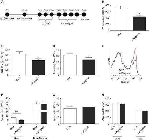
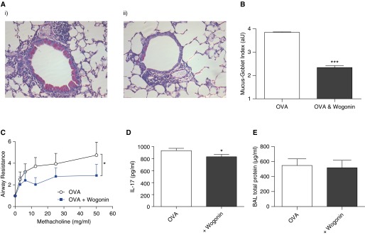
Methods: Human and mouse eosinophil apoptosis in response to wogonin was investigated by cellular morphology, flow cytometry, mitochondrial membrane permeability, and pharmacological caspase inhibition. Allergic lung inflammation was modeled in mice sensitized and challenged with ovalbumin. Bronchoalveolar lavage (BAL) and lung tissue were examined for inflammation, mucus production, and inflammatory mediator production. Airway hyperresponsiveness to aerosolized methacholine was measured.

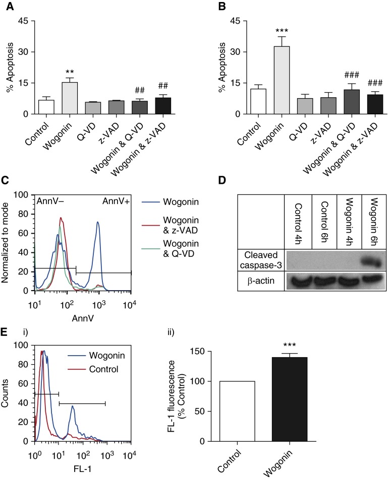
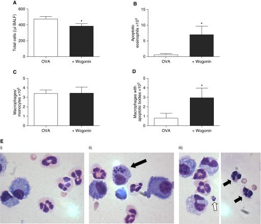
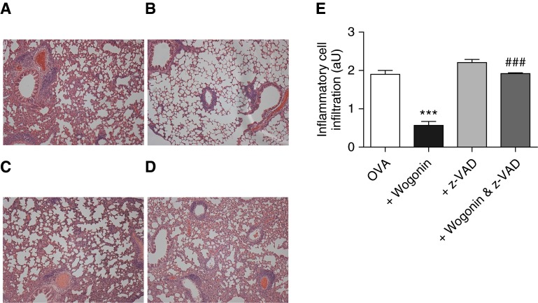
Measurements and main results: Wogonin induced time- and concentration-dependent human and mouse eosinophil apoptosis in vitro. Wogonin-induced eosinophil apoptosis occurred with activation of caspase-3 and was inhibited by pharmacological caspase inhibition. Wogonin administration attenuated allergic airway inflammation in vivo with reductions in BAL and interstitial eosinophil numbers, increased eosinophil apoptosis, reduced airway mucus production, and attenuated airway hyperresponsiveness. This wogonin-induced reduction in allergic airway inflammation was prevented by concurrent caspase inhibition in vivo.

Conclusions: Wogonin induces eosinophil apoptosis and attenuates allergic airway inflammation, suggesting that it has therapeutic potential for the treatment of allergic inflammation in humans.

Keywords: airway resistance; antiinflammatory agents; hypersensitivity; inflammation resolution; mucus.


Figures

Figure 1.
Figure 1.
Wogonin induces time- and concentration-dependent apoptosis of human eosinophils. (AC) Eosinophils were cultured for (A) 4–20 hours, (B) 6 hours, or (C) 20 hours with increasing concentrations of the flavone wogonin prior to eosinophil viability and apoptosis determination by flow cytometry (annexin V/propidium iodide [AnnV/PI] binding) (n ≥ 4). (D, E) Representative flow cytometry plots (x axis: AnnV; y axis: PI) and representative cytocentrifuge preparations (×1,000 original magnification) at 6 hours for (D) control and (E) 75 μM wogonin-treated eosinophils. Black arrow points to an eosinophil with apoptotic morphology. Data are expressed as mean ± SEM as analyzed by two-way analysis of variance (ANOVA) with a Bonferroni test (A) or one-way ANOVA with a Dunnett test (B, C). *P < 0.05, **P < 0.01, and ***P < 0.001 versus control.
Figure 2.
Figure 2.
Wogonin-induced eosinophil apoptosis is caspase dependent. Eosinophils were cultured for either (A) 6 hours or (B) 20 hours with the flavone wogonin (75 μM) with and without the broad-spectrum caspase inhibitors Q-VD (20 μM) or z-VAD (100 μM) prior to apoptosis determination by flow cytometry (annexin V/propidium iodide [AnnV/PI] binding) (n ≥ 5). (C) Representative overlay histogram of AnnV binding of eosinophils after 6 hours of treatment with wogonin (75 μM; blue line), wogonin plus z-VAD (100 μM; red line), and wogonin plus Q-VD (20 μM; green line). (D) Eosinophils were cultured for 4 hours or 6 hours in either control medium or wogonin (75 μM) before lysing and Western blotting for cleaved caspase-3 (17/19 kD) and β-actin (42 kD). (E) Eosinophils were cultured for 4 hours in either control medium or wogonin (100 μM) prior to analysis of mitochondrial membrane potential (Δψm), with loss of Δψm indicated by an increase in fluorescence channel (FL-1) fluorescence. A representative overlay histogram of FL-1 fluorescence is shown in (E, i), and cumulative data expressed relative to control are represented in (E, ii) (n = 7). Data are expressed as mean ± SEM as analyzed by analysis of variance with a Newman-Keuls multiple-comparisons test (A, B) or by t test (E). **P < 0.01 and ***P < 0.001 versus control; ##P < 0.01 and ###P < 0.001 versus wogonin-treated sample.
Figure 3.
Figure 3.
Mouse eosinophils recruited to sites of allergic inflammation undergo wogonin-induced apoptosis ex vivo. (A) Schema of the experimental protocol. (B) Representative flow cytometry plot (x axis: Siglec-F; y axis: Ly6G) demonstrating delineation of the eosinophil population (Ly6G−ve/Siglec-F+ve) as well as a smaller neutrophil population (Ly6G+ve/Siglec-F−ve). (C) Eosinophil apoptosis (AnnV+ve/DAPI−ve/Ly6G−ve/Siglec-F+ve) at 6 hours with increasing concentrations of wogonin (10–100 μM) with or without the caspase inhibitor Q-VD (20 μM) are shown (n = 3 or 4 separate experiments). (D) Bronchoalveolar lavage (BAL) cells incubated in control media or wogonin (100 μM) for 4 hours prior to lysing and Western blotting for cleaved caspase-3 (17/19 kD) and β-actin (42 kD). (E) Representative cytocentrifuge preparations (×1,000 original magnification) at 6 hours for (i) control cells and cells treated with (ii) wogonin (100 μM) and (iii) combined wogonin (100 μM) and Q-VD (20 μM). Black arrows indicate apoptotic eosinophils. Data are expressed as mean ± SEM as analyzed by analysis of variance with a Newman-Keuls multiple-comparisons test. *P < 0.05, **P < 0.01, ***P < 0.001 versus control, ###P < 0.001 versus 100 μM wogonin-treated sample. AnnV = annexin V; DAPI = 4′,6-diamidino-2-phenylindole; i.p. = intraperitoneal; i.t. = intratracheal; OVA = ovalbumin.
Figure 4.
Figure 4.
Wogonin attenuates allergic airway inflammation in vivo. (A) Following ovalbumin (OVA) sensitization and challenges, mice were treated with wogonin (1 mg) or vehicle control on Days 25–28, with acquisition and analysis of tissue taking place on Day 29. (B) Total bronchoalveolar lavage (BAL) cells, (C) BAL eosinophils (BAL eos), and (D) interstitial eosinophils are shown, with (E) representative overlay histogram of Siglec-F binding of lung interstitial inflammatory cells from mice treated with OVA (red line) or OVA + wogonin (blue line). (F) Blood and bone marrow eosinophils, (G) interstitial mast cells, and (H) interstitial and BAL eosinophil CD11b expression are shown. n = 7 or 8 mice. Data are expressed as mean ± SEM as analyzed by unpaired t test. *P <  0.05 versus vehicle. BALF = bronchoalveolar fluid; i.p. = intraperitoneal; i.t. = intratracheal; MFI = mean fluorescence intensity; n.s. = not significant.
Figure 5.
Figure 5.
Wogonin improves allergic airway responses. Following ovalbumin (OVA) sensitization and challenges, mice were treated with wogonin (1 mg) or vehicle control on Days 25–28, with acquisition and analysis of tissue and lung function performed on Day 29. (A) Representative lung tissue sections stained with periodic acid–Schiff stain in (i) OVA- and (ii) OVA- and wogonin-treated animals (×200 original magnification). (B) Quantification of mucus production was assessed by the mucus-goblet index, an average severity score per airway, expressed in arbitrary units (aU) (n = 4 or 5 mice per group). (C) Airway responsiveness was assessed in anesthetized and mechanically ventilated mice in response to aerosolized methacholine, with lung resistance determined and expressed relative to baseline values after phosphate-buffered saline exposure. (D) Lung IL-17 concentration and (E) bronchoalveolar lavage (BAL) protein (n ≥ 6). Data are expressed as mean ± SEM as analyzed by two-way analysis of variance (C) or unpaired t test (B, D, and E). *P < 0.05 and ***P < 0.001 versus control.
Figure 6.
Figure 6.
Wogonin accelerates eosinophil apoptosis in vivo. Following ovalbumin (OVA) sensitization and challenges, mice were treated with wogonin (1 mg) or vehicle control on Day 25, with acquisition and analysis of tissue performed on Day 26 (16 h after wogonin treatment). (A) Total bronchoalveolar lavage (BAL) inflammatory cells, (B) apoptotic eosinophils, (C) total macrophage/monocytes, and (D) macrophages containing apoptotic bodies (n ≥ 6). (E) BAL cells (×1,000 original magnification) demonstrate (i) normal eosinophil morphology in the control group, (ii) a macrophage containing apoptotic bodies in the wogonin-treated group (black arrow), and (iii) an apoptotic body (white arrow) and apoptotic eosinophils (black arrows) in the wogonin-treated group. Data are expressed as mean ± SEM, analyzed by unpaired t test. *P < 0.05 versus control. BALF = bronchoalveolar fluid.
Figure 7.
Figure 7.
Caspase inhibition prevents wogonin-induced attenuation of allergic airway inflammation. Following ovalbumin (OVA) sensitization and challenges, mice were treated with vehicle control, wogonin (1 mg at 32 h), z-VAD (5 mg/kg at 32, 36, 40, and 44 h), or combined wogonin and z-VAD, with acquisition and analysis of lungs for histology performed at 48 hours (Day 26). Representative lung tissue sections (hematoxylin and eosin stain) in (A) control/OVA, (B) wogonin-treated, (C) z-VAD-treated, and (D) combined wogonin- and z-VAD-treated animals (all images obtained at ×100 original magnification). (E) Quantification of interstitial and alveolar inflammatory cell infiltration, expressed in arbitrary units (aU) (n = 6 or 7 per group). Data are expressed as mean ± SEM, analyzed by analysis of variance with a Newman-Keuls multiple-comparison test. ***P < 0.001 versus control; ###P < 0.001 versus wogonin-treated mice.

Comment in

References

    1. Rosenberg HF, Dyer KD, Foster PS. Eosinophils: changing perspectives in health and disease. Nat Rev Immunol. 2013;13:9–22. - PMC - PubMed
    1. Fulkerson PC, Rothenberg ME. Targeting eosinophils in allergy, inflammation and beyond. Nat Rev Drug Discov. 2013;12:117–129. - PMC - PubMed
    1. Goh YP, Henderson NC, Heredia JE, Red Eagle A, Odegaard JI, Lehwald N, Nguyen KD, Sheppard D, Mukundan L, Locksley RM, et al. Eosinophils secrete IL-4 to facilitate liver regeneration. Proc Natl Acad Sci USA. 2013;110:9914–9919. - PMC - PubMed
    1. Lee JJ, Dimina D, Macias MP, Ochkur SI, McGarry MP, O’Neill KR, Protheroe C, Pero R, Nguyen T, Cormier SA, et al. Defining a link with asthma in mice congenitally deficient in eosinophils. Science. 2004;305:1773–1776. - PubMed
    1. Humbles AA, Lloyd CM, McMillan SJ, Friend DS, Xanthou G, McKenna EE, Ghiran S, Gerard NP, Yu C, Orkin SH, et al. A critical role for eosinophils in allergic airways remodeling. Science. 2004;305:1776–1779. - PubMed

Publication types