. 2015 Apr;156(4):711-721.
doi: 10.1097/j.pain.0000000000000103.

Dorsal root ganglion myeloid zinc finger protein 1 contributes to neuropathic pain after peripheral nerve trauma

Affiliations

Dorsal root ganglion myeloid zinc finger protein 1 contributes to neuropathic pain after peripheral nerve trauma

Zhisong Li et al. Pain. 2015 Apr.

Erratum in

Abstract

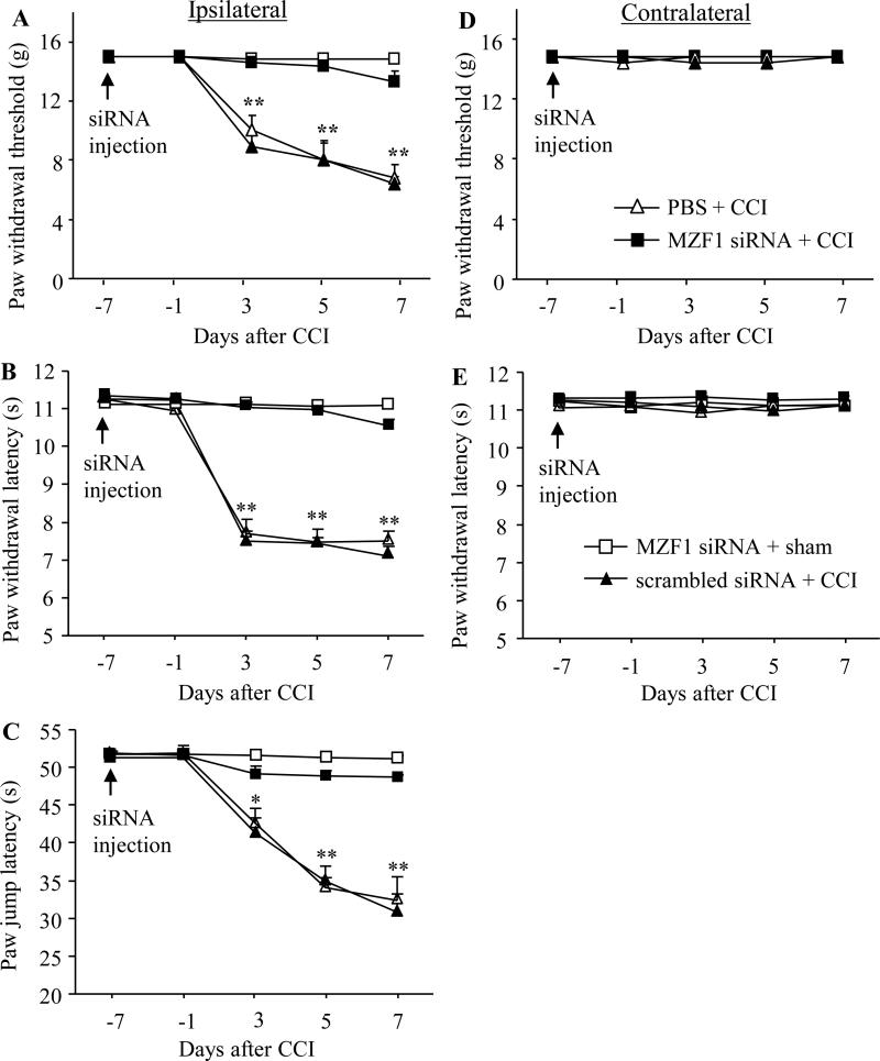
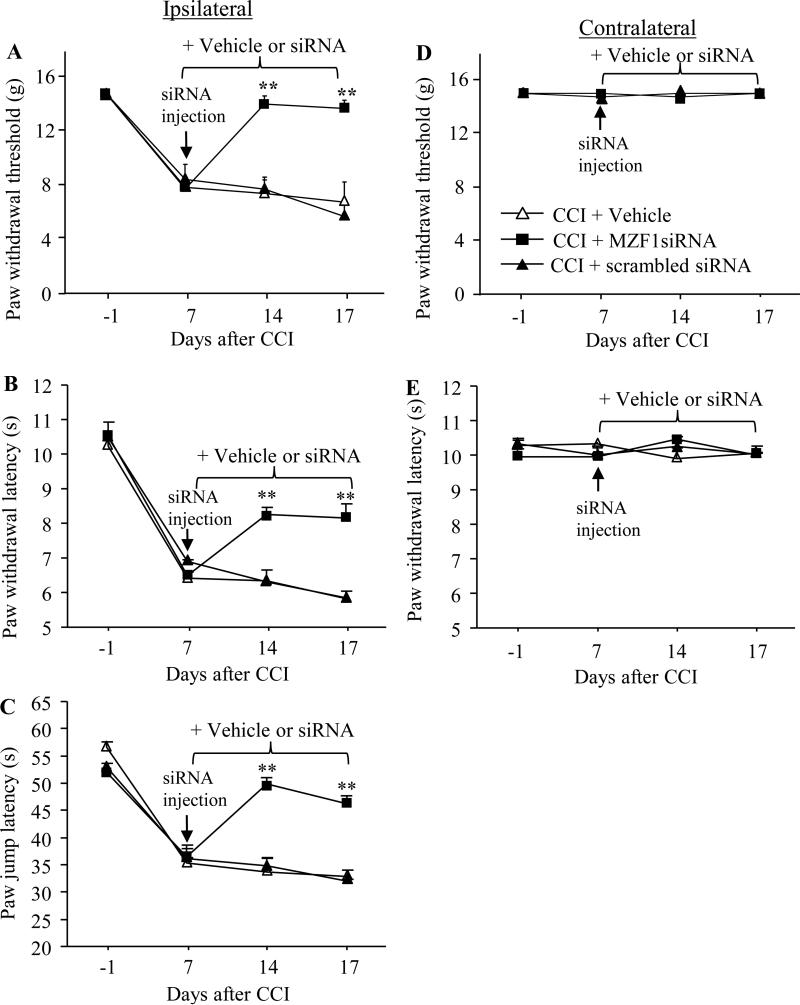
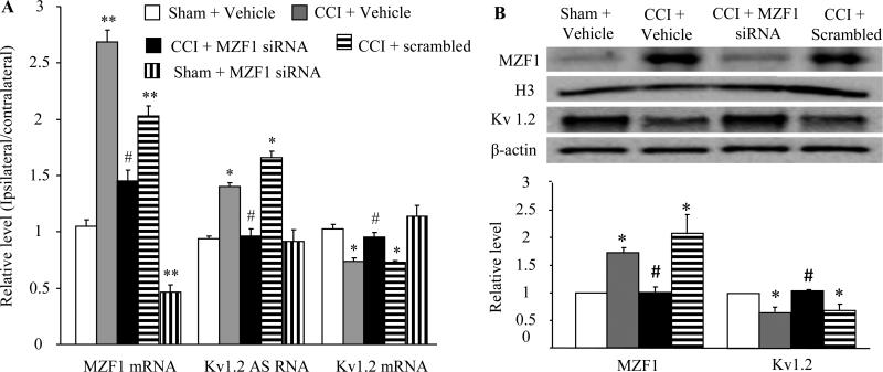
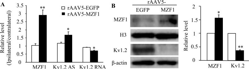
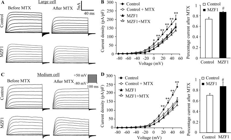
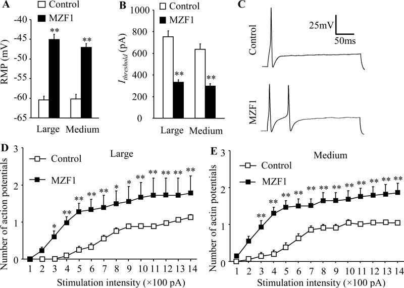
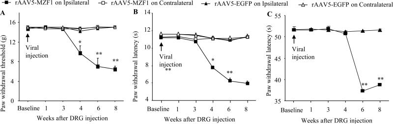
Peripheral nerve injury-induced changes in gene transcription and translation in primary sensory neurons of the dorsal root ganglion (DRG) are considered to contribute to neuropathic pain genesis. Transcription factors control gene expression. Peripheral nerve injury increases the expression of myeloid zinc finger protein 1 (MZF1), a transcription factor, and promotes its binding to the voltage-gated potassium 1.2 (Kv1.2) antisense (AS) RNA gene in the injured DRG. However, whether DRG MZF1 participates in neuropathic pain is still unknown. Here, we report that blocking the nerve injury-induced increase of DRG MZF1 through microinjection of MZF1 siRNA into the injured DRG attenuated the initiation and maintenance of mechanical, cold, and thermal pain hypersensitivities in rats with chronic constriction injury (CCI) of the sciatic nerve, without affecting locomotor functions and basal responses to acute mechanical, heat, and cold stimuli. Mimicking the nerve injury-induced increase of DRG MZF1 through microinjection of recombinant adeno-associated virus 5 expressing full-length MZF1 into the DRG produced significant mechanical, cold, and thermal pain hypersensitivities in naive rats. Mechanistically, MZF1 participated in CCI-induced reductions in Kv1.2 mRNA and protein and total Kv current and the CCI-induced increase in neuronal excitability through MZF1-triggered Kv1.2 AS RNA expression in the injured DRG neurons. MZF1 is likely an endogenous trigger of neuropathic pain and might serve as a potential target for preventing and treating this disorder.


Figures

Fig. 1
Fig. 1
Effect of pre-injection of MZF1 siRNA into the injured DRG on CCI-induced pain hypersensitivity during the development period. Pre-injection of MZF1 siRNA, but not scrambled siRNA, into the ipsilateral L4/5 DRGs abolished CCI-induced decreases in paw withdrawal threshold in response to mechanical stimuli (A), in paw withdrawal latency in responses to heat stimulation (B), and in paw jump latency in response to cold stimulation (C) on the ipsilateral side on days 3, 5, and 7 post-CCI. Neither MZF1 siRNA nor scrambled siRNA altered basal paw responses to mechanical (D) and heat (E) stimuli on the contralateral side of CCI rats during the observation period. No changes in basal paw responses were observed on either ipsilateral or contralateral side in sham rats injected with MZF1 siRNA (A-E). n = 8 rats/group. *P < 0.05 or **P < 0.01 vs the corresponding baseline.
Fig. 2
Fig. 2
Effect of post-injection of MZF1 siRNA into the injured DRG on CCI-induced pain hypersensitivity during the maintenance period. Microinjection of MZF1 siRNA, but not scrambled siRNA, into the ipsilateral L4/5 DRGs on day 7 post-CCI significantly reversed CCI-induced decreases in paw withdrawal threshold in response to mechanical stimuli (A), in paw withdrawal latency in responses to heat stimulation (B), and in paw jump latency in response to cold stimulation (C) on the ipsilateral side on days 14 and 17 post-CCI. Neither MZF1 siRNA nor scrambled siRNA altered basal paw responses to mechanical (D) and heat (E) stimuli on the contralateral side of CCI rats during the observation period. n = 5 rats/group. **P < 0.01 vs the CCI plus vehicle group at the corresponding time points.
Fig. 3
Fig. 3
Effect of microinjection of MZF1 siRNA into the injured DRG on the expression of MZF1 mRNA and protein, Kv1.2 AS RNA, and Kv1.2 mRNA and protein in the injured DRG on day 7 post-CCI or sham surgery. (A) CCI increased the expression of MZF1 mRNA and Kv1.2 AS RNA and decreased the expression of Kv1.2 mRNA in the ipsilateral L4/5 DRGs. These effects were significantly blocked by pre-injection of MZF1 siRNA, but not scrambled siRNA. The level of MZF1 mRNA was reduced in the sham rats pre-injected with MZF1 siRNA. (B) CCI increased the expression of MZF1 protein and decreased the expression of Kv1.2 protein the ipsilateral L4/5 DRGs. These effects were markedly blocked by pre-injection of MZF1 siRNA, but not scrambled siRNA. *P < 0.05, **P < 0.01 vs the corresponding sham plus vehicle group. n = 3 rats/assay. #P < 0.05 vs the corresponding CCI plus vehicle group.
Fig. 4
Fig. 4
Effect of microinjection of rAAV5-MZF1 into the DRG on the expression of MZF1 mRNA and protein, Kv1.2 AS RNA, and Kv1.2 mRNA and protein in the DRG of naïve rats. (A) Injection of rAAV5-MZF1 into unilateral L4/5 DRGs produced increases in the levels of MZF1 mRNA and Kv1.2 AS RNA and a decrease in the amount of Kv1.2 mRNA in the ipsilateral L4/5 DRGs. (B) Injection of rAAV5-MZF1 into unilateral L4/5 DRGs produced an increase in the level of MZF1 protein and a decrease in the amount of Kv1.2 protein in the ipsilateral L4/5 DRGs. N = 3 rats/assay. *P < 0.05, **P < 0.01 vs the corresponding AAV5-EGFP group.
Fig. 5
Fig. 5
Effect of microinjection of rAAV5-MZF1 into the DRG on total Kv current in large and medium DRG neurons. (A) Representative traces of total Kv current in large DRG neurons from control (rAAV5-EGFP)- and MZF1(rAAV5-MZF1 plus rAAV5-EGFP)-treated rats before or after bath perfusion of 100 nM maurotoxin (MTX). (B) Left: I-V curve for control- and MZF1-treated large DRG neurons before or after 100 nM MTX treatment. The current density was plotted against each step testing voltage. Right: at +50 mV, the reduction in total Kv current after MTX treatment in large DRG neurons was greater in the control group than in the MZF1-treated group. n = 11 cells for control group (8 DRGs from 4 rats) and n = 14 for MZF1-treated group (8 DRGs from 4 rats). (C) Representative traces of total Kv current in medium DRG neurons from control- and MZF1-treated rats before or after bath perfusion of 100 nM MTX. (D) Left: I-V curve for control- and MZF1-treated medium DRG neurons before or after 100 nM MTX treatment. The current density was plotted against each step testing voltage. Right: at +50 mV, the reduction in total Kv current after MTX treatment in medium DRG neurons was greater in the control group (8 DRGs from 4 rats) than in the MZF1-treated group (8 DRGs from 4 rats). n = 17 cells for control group and n = 16 for MZF1-treated group. *P < 0.05, **P < 0.1 vs the corresponding the MZF1-treated group. #P < 0.05 vs the corresponding control group.
Fig. 6
Fig. 6
Effect of microinjection of rAAV5-MZF1 into the DRG on excitability in large and medium DRG neurons. n = 24 large, 21 medium for control group (8 DRGs from 4 rats) and n = 18 large, 28 medium for MZF1-treated group (8 DRGs from 4 rats). (A, B) Resting membrane potential (RMP; A) and current threshold for pulses (Ithreshold; B). **P < 0.01 vs the corresponding control group. (C) Representative traces of the evoked action potentials in large DRG neurons. (D, E) Numbers of evoked action potentials from large (D) and medium (E) DRG neurons of control and MZF1-treated rats after application of different currents. *P < 0.05, **P < 0.01 vs the corresponding same stimulation intensity in the control group.
Fig. 7
Fig. 7
Effect of microinjection of rAAV5-MZF1 into the DRG on nociceptive threshold in naïve rats. Paw withdrawal responses to mechanical (A), heat (B), and cold (C) stimuli from the rAAV5-EGFP-injected (n = 6 rats) and rAAV5-MZF1-injected (n = 5 rats) groups. *P < 0.05, **P < 0.01 vs the rAAV5-EGFP-injected group on the ipsilateral side at the corresponding time points.

References

    1. Abrahamsen B, Zhao J, Asante CO, Cendan CM, Marsh S, Martinez-Barbera JP, Nassar MA, Dickenson AH, Wood JN. The cell and molecular basis of mechanical, cold, and inflammatory pain. Science. 2008;321:702–705. - PubMed
    1. Amaya F, Wang H, Costigan M, Allchorne AJ, Hatcher JP, Egerton J, Stean T, Morisset V, Grose D, Gunthorpe MJ, Chessell IP, Tate S, Green PJ, Woolf CJ. The voltage-gated sodium channel Na(v)1.9 is an effector of peripheral inflammatory pain hypersensitivity. J Neurosci. 2006;26:12852–12860. - PMC - PubMed
    1. Bennett GJ, Xie YK. Af peripheral mononeuropathy in rat that produces disorders of pain sensation like those seen in man. Pain. 1988;33:87–107. - PubMed
    1. Campbell JN, Meyer RA. Mechanisms of neuropathic pain. Neuron. 2006;52:77–92. - PMC - PubMed
    1. Castle NA, London DO, Creech C, Fajloun Z, Stocker JW, Sabatier JM. Maurotoxin: a potent inhibitor of intermediate conductance Ca2+-activated potassium channels. Mol Pharmacol. 2003;63:409–418. - PubMed

Publication types

MeSH terms