. 2015 Apr;156(4):722-730.
doi: 10.1097/j.pain.0000000000000104.

Sensitization of nociceptive spinal neurons contributes to pain in a transgenic model of sickle cell disease

Affiliations

Sensitization of nociceptive spinal neurons contributes to pain in a transgenic model of sickle cell disease

Giuseppe Cataldo et al. Pain. 2015 Apr.

Abstract

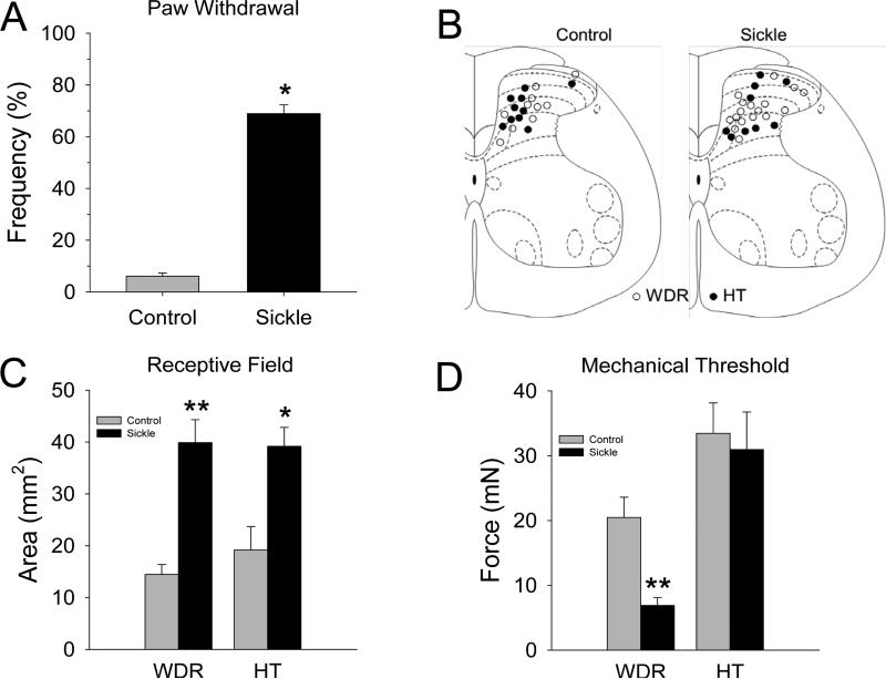
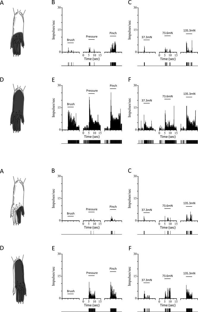
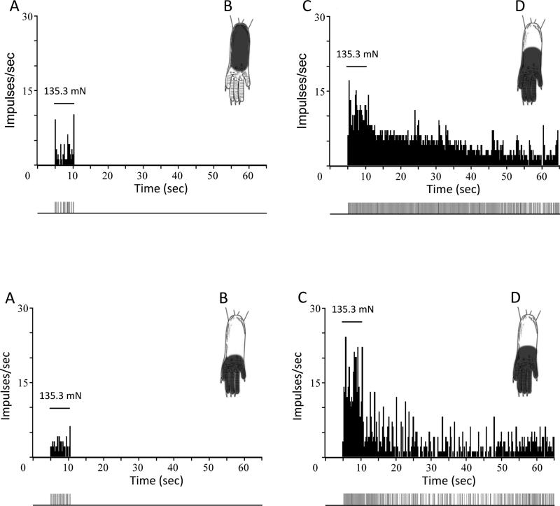
Chronic pain is a major characteristic feature of sickle cell disease (SCD). The refractory nature of pain and the development of chronic pain syndromes in many patients with SCD suggest that central neural mechanisms contribute to pain in this disease. We used HbSS-BERK sickle mice, which show chronic features of pain similar to those observed in SCD, and determined whether sensitization of nociceptive neurons in the spinal cord contributes to pain and hyperalgesia in SCD. Electrophysiological recordings of action potential activity were obtained from single identified dorsal horn neurons of the spinal cord in anesthetized mice. Compared with control HbAA-BERK mice, nociceptive dorsal horn neurons in sickle mice exhibited enhanced excitability as evidenced by enlarged receptive fields, increased rate of spontaneous activity, lower mechanical thresholds, enhanced responses to mechanical stimuli, and prolonged afterdischarges following mechanical stimulation. These changes were accompanied by increased phosphorylation of mitogen-activated protein kinases (MAPKs) in the spinal cord that are known to contribute to neuronal hyperexcitability, including c-Jun N-terminal kinase (JNK), p44/p42 extracellular signaling-regulated kinase (ERK), and p38. These findings demonstrate that central sensitization contributes to pain in SCD.


Figures

Figure 1
Figure 1
A) Sickle mice exhibited mechanical hyperalgesia. None of the control mice (n=14) exhibited a withdrawal response frequency greater than 15% (range: 0 to 15%) to this force, whereas sickle mice (n=14) had response frequencies that ranged between 50-100%. The mean (±SEM) frequency of paw withdrawal differed between the groups (t-test, p<0.001). B) Schematic representation of the location of recording sites in the spinal dorsal horn. The location of recording sites for WDR and HT neurons were similar for control (left) and sickle (right) mice and were located throughout the dorsal horn. The mean (±SEM) depths of recording sites from the surface of the spinal cord did not differ between control and sickle mice (307.1 ±32.6 μm and 399.1 ±33.3 μm, respectively). C) The mechano-sensitive receptive field areas of dorsal horn neurons differed between control and sickle mice. Receptive field areas were larger for both WDR and HT neurons in sickle mice (t-tests; *P < 0.05; **P < 0.01). D) Mechanical thresholds of WDR neurons were lower in mice with SCD as compared to controls (t-tests; **P < 0.01).
Figure 2
Figure 2
Receptive field (RF) areas and responses of nociceptive neurons evoked by mechanical stimuli are greater in sickle mice. Upper panels: Location of the RF, and peristimulus time histograms showing discharge rates evoked by stimuli used for functional characterization (brush, pressure pinch applied to the RF) and responses evoked by von Frey monofilaments of controlled force for a single WDR neuron from a control (top panels A, B, and C) and from a sickle (lower panels D, E, and F) mouse. Solid horizontal lines represent the time of application of the stimuli (5 s). Discriminated output pulses from a window discriminator are provided below the histograms. Lower panels: Same format as above but RF areas and evoked responses are shown for single HT neurons from a control (A-C) and sickle (D-F) mouse. Bin width for all peristimulus time histograms is 100 ms.
Figure 3
Figure 3
Mean (±SE) discharge rates (impulses/s) for all WDR (A) and HT (B) neurons evoked by application of 37.3 mN, 73.6 mN and 135.3 mN von Frey filaments in control and sickle mice. Responses evoked by the 73.6 mN and 135.3 mN stimuli were greater for WDR neurons in sickle mice whereas responses to each of the forces was greater in sickle mice. *P < 0.05; **P < 0.01).
Figure 4
Figure 4
Mechanical stimulation evokes prolonged afterdischarges in nociceptive neurons in sickle mice. Upper panels show responses evoked by the 135.3 mN force in a WDR neuron from a control mouse (A) and from a sickle mouse (B). Similarly, responses of single HT neurons from a control and from a sickle mouse are shown in C and D. Horizontal lines represent the time of stimulus application. The location of the RF is shown for each neuron. Discriminated output pulses from a window discriminator are provided below the histograms. Responses to mechanical stimulation were greater and prolonged in sickle mice. Bin width of the peristimulus time histograms is 250 ms.
Figure 5
Figure 5. Increased nociceptive signaling in the spinal cord of sickle mice
Protein bands (and molecular weight) of phosphorylated and total proteins determined using Western immunoblotting and their densitometric profiles are shown for JNK (A), p38 (B), and p44/p42 ERK (C). Density of phosphoprotein bands relative to their specific total protein are shown. Data are expressed as mean ± SEM from 5 mice per group of 5 separate experiments. Statistical significance is denoted by *P < 0.05 and **P < 0.005. Mean age of mice ± SEM in months were 4.04 ± 0.29 for HbAA-BERK and 4.38 ± 0.32 for HbSS-BERK.

References

    1. Amjad H, Bannerman RM, Judisch JM. Letter: sickling pain and season. Br Med J. 1974;2:54. - PMC - PubMed
    1. Ballas SK, Gupta K, Adams-Graves P. Sickle cell pain: a critical reappraisal. Blood. 2012;120:3647–56. - PubMed
    1. Bardoni R, Takazawa T, Tong CK, Choudhury P, Scherrer G, Macdermott AB. Pre- and postsynaptic inhibitory control in the spinal cord dorsal horn. Ann N Y Acad Sci. 2013;1279:90–6. - PMC - PubMed
    1. Brandow AM, Stucky CL, Hillery CA, Hoffmann RG, Panepinto JA. Patients with sickle cell disease have increased sensitivity to cold and heat. Am J Hematol. 2013;88:37–43. - PMC - PubMed
    1. Cain DM, Vang D, Simone DA, Hebbel RP, Gupta K. Mouse models for studying pain in sickle disease: effects of strain, age, and acuteness. Br J Haematol. 2012;156:535–44. - PMC - PubMed

Publication types

MeSH terms

Substances