. 2015 May;70(5):1367-80.
doi: 10.1093/jac/dku545. Epub 2015 Jan 27.

ST-246 is a key antiviral to inhibit the viral F13L phospholipase, one of the essential proteins for orthopoxvirus wrapping

Affiliations

ST-246 is a key antiviral to inhibit the viral F13L phospholipase, one of the essential proteins for orthopoxvirus wrapping

Sophie Duraffour et al. J Antimicrob Chemother. 2015 May.

Abstract

Objectives: ST-246 is one of the key antivirals being developed to fight orthopoxvirus (OPV) infections. Its exact mode of action is not completely understood, but it has been reported to interfere with the wrapping of infectious virions, for which F13L (peripheral membrane protein) and B5R (type I glycoprotein) are required. Here we monitored the appearance of ST-246 resistance to identify its molecular target.

Methods: Vaccinia virus (VACV), cowpox virus (CPXV) and camelpox virus (CMLV) with reduced susceptibility to ST-246 were selected in cell culture and further characterized by antiviral assays and immunofluorescence. A panel of recombinant OPVs was engineered and a putative 3D model of F13L coupled with molecular docking was used to visualize drug-target interaction. The F13L gene of 65 CPXVs was sequenced to investigate F13L amino acid heterogeneity.

Results: Amino acid substitutions or insertions were found in the F13L gene of six drug-resistant OPVs and production of four F13L-recombinant viruses confirmed their role(s) in the occurrence of ST-246 resistance. F13L, but not B5R, knockout OPVs showed resistance to ST-246. ST-246 treatment of WT OPVs delocalized F13L- and B5R-encoded proteins and blocked virus wrapping. Putative modelling of F13L and ST-246 revealed a probable pocket into which ST-246 penetrates. None of the identified amino acid changes occurred naturally among newly sequenced or NCBI-derived OPV F13L sequences.

Conclusions: Besides demonstrating that F13L is a direct target of ST-246, we also identified novel F13L residues involved in the interaction with ST-246. These findings are important for ST-246 use in the clinic and crucial for future drug-resistance surveillance programmes.

Keywords: antivirals; drug resistance; mechanisms of action; poxvirus; vaccinia virus.


Figures

Figure 1.
Figure 1.
Schematic overview of the amino acid changes identified in F13L of VACV-Cop, CPXV-BR and CMLV strains grown under ST-246 selective pressure. The amino acid changes in each virus are specified with respect to the reference virus for each species. The cell line in which the selection procedure was done is given in brackets.
Figure 2.
Figure 2.
Phenotyping of F13L-mutated VACV-Cop, CPXV-BR and CMLV in HEL and Vero cells. Two or three clones of each WT and ST-246R clone were used in plaque reduction assays and at least three to five independent experiments were performed per clone and per test compound (ST-246 and cidofovir). Assays were performed in HEL (left-hand panels) and Vero (right-hand panels) cells for the drug-resistant viruses selected in HEL and Vero cells, respectively (refer to Table 1 for names). The data are presented as a dot plot of the EC50s of ST-246R clones (open symbols) versus the EC50s of WT parent clones (filled symbols). On each graph are shown the fold changes in ST-246 EC50 concentrations, which were calculated by dividing the mean of EC50s for ST-246R clones by the mean of EC50s for WT clones. Results are presented as mean ± SD.
Figure 3.
Figure 3.
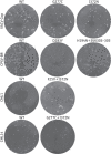
Plaque phenotype under liquid overlay. BSC-1 cells were infected with 25–50 pfu of the indicated strains. At day 3 post-infection, cell monolayers were fixed and stained with crystal violet.
Figure 4.
Figure 4.
Profiles of the susceptibility of F13L and B5R knockout OPVs to ST-246. (a) Drug-susceptibility profiles of WT, ΔF13L and ΔB5R clones in Vero cells. One clone of each stock was used in CPE reduction assays with ST-246, and at least three independent experiments were performed. The results are presented as a dot plot of the EC50s (mean ± SD). EC50s >26.5 µM were obtained with the two ΔF13L viruses. The fold change in ST-246 EC50 values was calculated as the ratio of mean EC50 for ΔF13L to mean EC50 for WT; it was >44 167-fold for VACV-Cop-ΔF13L (EC50 >26.5 µM) versus VACV-Cop (EC50 = 0.0006 ± 0.00007 µM) and >98-fold for CPXV-BR-ΔF13L (EC50 >26.5 µM) versus CPXV-BR (EC50 = 0.27 ± 0.17 µM). (b) Virus yields. Confluent Vero cells were infected with the indicated virus at an moi of 0.01 pfu per cell for 2 h, after which serial concentrations of ST-246 (µM) were added for 2 days. Extracellular (supernatant) and intracellular (cell-associated) fractions were collected and virus titres determined. Three independent experiments were performed and the data are presented as mean ± SD.
Figure 5.
Figure 5.
Intracellular localization of F13L and B5R in cells infected with WT viruses in the absence or presence of ST-246. BHK-21 cells infected with VACV-Cop, VACV-WR or CML1 for 18 h at an moi of 5 pfu per cell in the absence or presence of ST-246 (5 µg/mL) were subjected to immunofluorescence analysis. DNA was stained by incubation with Hoechst dye for 30 min at the end of the infection period. F13L was stained with rat anti-F13 antibody and B5R with rat anti-B5R antibody, both followed by donkey anti-rat Alexa Fluor 594 antibody. Note the delocalization of F13L and B5R upon treatment with ST-246, as evidenced by Golgi complexes appearing in fragmented vesicles, a diffuse cytoplasmic staining of F13L (especially marked with VACV-WR and CML1) and the absence of the spotted staining normally corresponding to virus particles in cells and cell projections. Inserts show zoom-ins of squared regions.
Figure 6.
Figure 6.
Intracellular distribution of F13L and plaque size in cells infected with WT or F13L-mutated CPXV-BR viruses in the absence or presence of ST-246. BSC-1 (plaque size) or BHK-21 (immunofluorescence) cells were infected with the indicated virus for, respectively, 3 days (moi 100 pfu per cell) or 18 h (moi 5 pfu per cell) in the absence or presence of ST-246 (5 µg/mL). To determine plaque size, cell monolayers were fixed in ethanol and stained with crystal violet and photographs were taken. To assess immunofluorescence, DNA was stained by incubation with Hoechst dye for 30 min at the end of the infection period. F13L was stained with rat anti-F13 antibody followed by donkey anti-rat Alexa Fluor 594 antibody. Inserts show zoom-ins of squared regions.
Figure 7.
Figure 7.
F13L modelling and molecular docking of F13L/ST-246. The structure of Streptomyces sp. (strain PMF) phospholipase D (code pdb 1V0W) was used to build a putative model of the F13L protein. (a) Overview of the F13L putative 3D structure highlighting the residues important for ST-246 and/or IMCBH antiviral efficacy. The insert corresponds to the region defined for molecular docking. The putative hydrophobic region (172–198) that anchors F13L in cellular membranes is shown in yellow. (b) Docking of F13L with ST-246 or IMCBH revealing the presence of a pocket in which both of the molecules fit. (c) Close-up view of interaction site between F13L and ST-246 formed by G277, W279, D280, A288, A289, A290, L302, S303, V304, K305, V306 and F307. Note that the three fluorine atoms penetrate the pocket, while the benzamide moiety locates at the interface formed by W279 and V304. (d) Surface view (left-hand panel) and close-up view (right-hand panel) of the H(N)KD motif located opposite to the ST-246 interaction site. Binding of ST-246 may directly interfere with the flexibility of the chain bearing N312 and K314, which form the phospholipase motif. Red, residues found to be linked with ST-246 resistance and IMCBH resistance; cyan (A290), the change A290V was identified in a patient that received ST-246 therapy; orange and grey in (c), additional residues forming the interaction site of the inhibitor; yellow (N312), blue (K314) and green (D319), residues corresponding to the H(N)KD phospholipase motif; magenta (S365), residue corresponding to the last amino acid (S365) that could be integrated in the modelling. The last 7 amino acids of F13L (372 amino acids) could not be included in the modelling.

Similar articles

Cited by

References

    1. Shchelkunov SN. An increasing danger of zoonotic orthopoxvirus infections. PLoS Pathog 2013; 9: e1003756. - PMC - PubMed
    1. Favier AL, Flusin O, Lepreux S, et al. Necrotic ulcerated lesion in a young boy caused by cowpox virus infection. Case Rep Dermatol 2011; 3: 186–94. - PMC - PubMed
    1. Haase O, Moser A, Rose C, et al. Generalized cowpox infection in a patient with Darier disease. Br J Dermatol 2011; 164: 1116–8. - PubMed
    1. Pelkonen PM, Tarvainen K, Hynninen A, et al. Cowpox with severe generalized eruption, Finland. Emerg Infect Dis 2003; 9: 1458–61. - PMC - PubMed
    1. Vogel S, Sardy M, Glos K, et al. The Munich outbreak of cutaneous cowpox infection: transmission by infected pet rats. Acta Derm Venereol 2012; 92: 126–31. - PubMed

Publication types

MeSH terms