

Measuring Neuromuscular Junction Functionality in the SOD1(G93A) Animal Model of Amyotrophic Lateral Sclerosis
- PMID: 25631208
- PMCID: PMC4516896
- DOI: 10.1007/s10439-015-1259-x
Measuring Neuromuscular Junction Functionality in the SOD1(G93A) Animal Model of Amyotrophic Lateral Sclerosis
Abstract
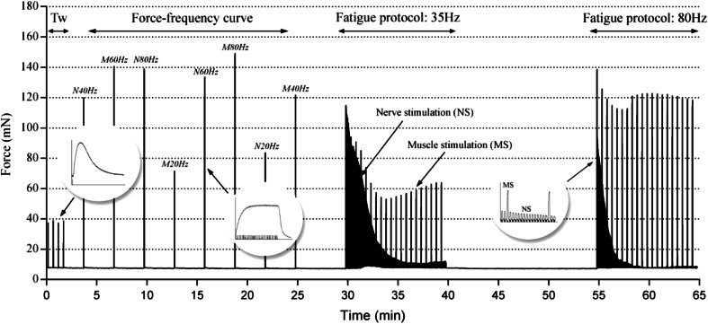
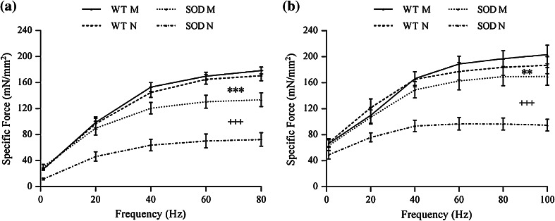
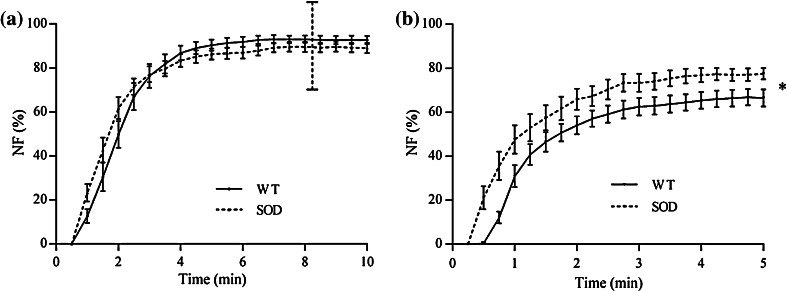
Amyotrophic lateral sclerosis (ALS) is a fatal neurodegenerative disease that leads to motor neuron degeneration, alteration in neuromuscular junctions (NMJs), muscle atrophy, and paralysis. To investigate the NMJ functionality in ALS we tested, in vitro, two innervated muscle types excised from SOD1(G93A) transgenic mice at the end-stage of the disease: the Soleus, a postural muscle almost completely paralyzed at that stage, and the diaphragm, which, on the contrary, is functional until death. To this aim we employed an experimental protocol that combined two types of electrical stimulation: the direct stimulation and the stimulation through the nerve. The technique we applied allowed us to determine the relevance of NMJ functionality separately from muscle contractile properties in SOD1(G93A) animal model. Functional measurements revealed that the muscle contractility of transgenic diaphragms is almost unaltered in comparison to control muscles, while transgenic Soleus muscles were severely compromised. In contrast, when stimulated via the nerve, both transgenic muscle types showed a strong decrease of the contraction force, a slowing down of the kinetic parameters, as well as alterations in the neurotransmission failure parameter. All together, these results confirm a severely impaired functionality in the SOD1(G93A) neuromuscular junctions.
Figures


Similar articles
-
Macrophage-mediated inflammation and glial response in the skeletal muscle of a rat model of familial amyotrophic lateral sclerosis (ALS).Exp Neurol. 2016 Mar;277:275-282. doi: 10.1016/j.expneurol.2016.01.008. Epub 2016 Jan 13. Exp Neurol. 2016. PMID: 26775178 Free PMC article.
-
AAV-NRIP gene therapy ameliorates motor neuron degeneration and muscle atrophy in ALS model mice.Skelet Muscle. 2024 Jul 24;14(1):17. doi: 10.1186/s13395-024-00349-z. Skelet Muscle. 2024. PMID: 39044305 Free PMC article.
-
Progressive impairment of CaV1.1 function in the skeletal muscle of mice expressing a mutant type 1 Cu/Zn superoxide dismutase (G93A) linked to amyotrophic lateral sclerosis.Skelet Muscle. 2016 Jun 23;6:24. doi: 10.1186/s13395-016-0094-6. eCollection 2016. Skelet Muscle. 2016. PMID: 27340545 Free PMC article.
-
Neuromuscular junction destruction during amyotrophic lateral sclerosis: insights from transgenic models.Curr Opin Pharmacol. 2009 Jun;9(3):341-6. doi: 10.1016/j.coph.2009.03.007. Epub 2009 Apr 20. Curr Opin Pharmacol. 2009. PMID: 19386549 Review.
-
Unraveling the complexity of amyotrophic lateral sclerosis: recent advances from the transgenic mutant SOD1 mice.CNS Neurol Disord Drug Targets. 2010 Aug;9(4):491-503. doi: 10.2174/187152710791556096. CNS Neurol Disord Drug Targets. 2010. PMID: 20522008 Review.
Cited by
-
Repeated Measurement of Respiratory Muscle Activity and Ventilation in Mouse Models of Neuromuscular Disease.J Vis Exp. 2017 Apr 17;(122):55599. doi: 10.3791/55599. J Vis Exp. 2017. PMID: 28448001 Free PMC article.
-
Breathing: Motor Control of Diaphragm Muscle.Physiology (Bethesda). 2018 Mar 1;33(2):113-126. doi: 10.1152/physiol.00002.2018. Physiology (Bethesda). 2018. PMID: 29412056 Free PMC article. Review.
-
Development of a Novel Technique for the Measurement of Neuromuscular Junction Functionality in Isotonic Conditions.Cell Mol Bioeng. 2022 Apr 7;15(3):255-265. doi: 10.1007/s12195-022-00721-3. eCollection 2022 Jun. Cell Mol Bioeng. 2022. PMID: 35611165 Free PMC article.
-
Mechanisms of compensatory plasticity for respiratory motor neuron death.Respir Physiol Neurobiol. 2019 Jul;265:32-39. doi: 10.1016/j.resp.2019.01.001. Epub 2019 Jan 6. Respir Physiol Neurobiol. 2019. PMID: 30625378 Free PMC article. Review.
-
An Optimized Ex Vivo Protocol for Quantitative Electrophysiological Assessment of Neuromuscular Junctions and Skeletal Muscle Function Using the Aurora System.Bio Protoc. 2025 Jun 20;15(12):e5353. doi: 10.21769/BioProtoc.5353. eCollection 2025 Jun 20. Bio Protoc. 2025. PMID: 40625426 Free PMC article.
References
-
- Aldrich TK, Shander A, Chaudhry I, Nagashima H. Fatigue of isolated rat diaphragm: role of impaired neuromuscular transmission. J. Appl. Physiol. 1986;61:1077–1083. - PubMed
-
- Asmussen G, Gaunitz U. Temperature effects on isometric contractions of slow and fast twitch muscles of various rodents–dependence on fibre type composition: a comparative study. Biomed. Biochim. Acta. 1989;48:S536–S541. - PubMed
-
- Augusto V, Padovani CR, Compos GER. Skeletal muscle fiber types in C57BL6J mice. Braz. J. morphol. Sci. 2004;21:89–94.
Publication types
MeSH terms
Substances
Grants and funding
LinkOut - more resources
Full Text Sources
Other Literature Sources
Medical
Miscellaneous
