Review
. 2015 Mar;79(1):1-17.
doi: 10.1128/MMBR.00041-14.

Yeast prions: structure, biology, and prion-handling systems

Affiliations
Review

Yeast prions: structure, biology, and prion-handling systems

Reed B Wickner et al. Microbiol Mol Biol Rev. 2015 Mar.

Abstract

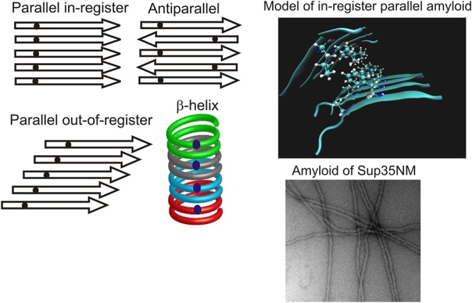
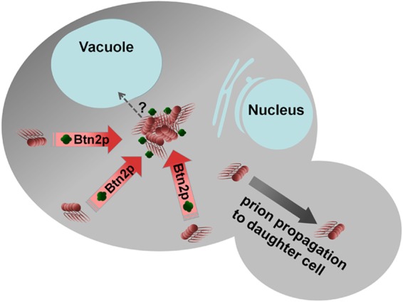
A prion is an infectious protein horizontally transmitting a disease or trait without a required nucleic acid. Yeast and fungal prions are nonchromosomal genes composed of protein, generally an altered form of a protein that catalyzes the same alteration of the protein. Yeast prions are thus transmitted both vertically (as genes composed of protein) and horizontally (as infectious proteins, or prions). Formation of amyloids (linear ordered β-sheet-rich protein aggregates with β-strands perpendicular to the long axis of the filament) underlies most yeast and fungal prions, and a single prion protein can have any of several distinct self-propagating amyloid forms with different biological properties (prion variants). Here we review the mechanism of faithful templating of protein conformation, the biological roles of these prions, and their interactions with cellular chaperones, the Btn2 and Cur1 aggregate-handling systems, and other cellular factors governing prion generation and propagation. Human amyloidoses include the PrP-based prion conditions and many other, more common amyloid-based diseases, several of which show prion-like features. Yeast prions increasingly are serving as models for the understanding and treatment of many mammalian amyloidoses. Patients with different clinical pictures of the same amyloidosis may be the equivalent of yeasts with different prion variants.


Figures

FIG 1
FIG 1
Prions [URE3], [PSI+], and [PIN+] of S. cerevisiae and [Het-s] of Podospora anserina. These prions are based on self-propagating amyloids of Ure2p, Sup35p, Rnq1p, and HET-s, respectively. The prion domains of Ure2p and Sup35p have nonprion functions, explaining their retention in evolution despite detrimental prion formation.
FIG 2
FIG 2
[BETA] prion based on autocatalysis of Prb1p activation. Prb1p (vacuolar protease B) is made as an inactive precursor which can be activated by the active form of the same protein (67). Thus, a cell starting with no active enzyme remains so, while a cell with active enzyme continues to activate the precursor as it is synthesized. Transmission of active Prb1p to a cell lacking the active form “infects” it with the [BETA] prion (68). The active form of protease B (red) can cleave inactive (blue) precursor molecules (at sites labeled “autocleavage”) to activate them.
FIG 3
FIG 3
β-Helix versus in-register parallel β-sheet for amyloid structure. (Left) Four types of β-sheet. Small dark dots represent a single 13C-labeled atom in each protein molecule. Only for the in-register parallel architecture will the labeled atoms have an ∼0.5-nm spacing, while the spacing (measured by solid-state NMR) will be much greater for the other structures. (Top right) Model of an in-register parallel structure based on data from reference . The only side chains shown are those of a single residue in the top sheet and a single residue in the bottom sheet. (Bottom right) Electron microscopic image of amyloid formed from recombinant Sup35NM. Magnification, ×56,000.
FIG 4
FIG 4
Proposed mechanism of conformational templating by prion protein amyloids. Energetically favorable interactions between identical side chains enforce the in-register architecture of these amyloids. H-bonds between the side chains of identical Gln residues, for example, can form only if the residues are aligned in register. Interactions between charged side chains would be unfavorable, and charged side chains are rare in yeast prion domains. Similarly, in order to form these favorable interactions, a new molecule being added to the end of the filament must assume the same conformation as that of molecules previously added to the filament. Thus, the protein can template its own conformation, just as a DNA or RNA can template its own sequence (7, 216).
FIG 5
FIG 5
Mechanism of seed generation by Hsp104-Hsp70-Hsp40. Hsp104, working with Hsp40 (mainly Sis1p) and Hsp70 (Ssa proteins), extracts a monomer from an amyloid filament, resulting in the formation of two filaments, with two more growing ends. (Adapted from reference .)
FIG 6
FIG 6
Model of Btn2 curing of the [URE3] prion. Btn2p sequesters amyloid filaments at a single site, increasing the probability of prion loss on cell division (96, 187).

References

    1. Alper T, Haig DA, Clarke MC. 1966. The exceptionally small size of the scrapie agent. Biochem Biophys Res Commun 22:278–284. doi:10.1016/0006-291X(66)90478-5. - DOI - PubMed
    1. Griffith JS. 1967. Self-replication and scrapie. Nature 215:1043–1044. doi:10.1038/2151043a0. - DOI - PubMed
    1. Wickner RB. 1994. [URE3] as an altered URE2 protein: evidence for a prion analog in S. cerevisiae. Science 264:566–569. doi:10.1126/science.7909170. - DOI - PubMed
    1. Baxa U, Wickner RB, Steven AC, Anderson D, Marekov L, Yau W-M, Tycko R. 2007. Characterization of β-sheet structure in Ure2p1-89 yeast prion fibrils by solid state nuclear magnetic resonance. Biochemistry 46:13149–13162. doi:10.1021/bi700826b. - DOI - PubMed
    1. Shewmaker F, Wickner RB, Tycko R. 2006. Amyloid of the prion domain of Sup35p has an in-register parallel β-sheet structure. Proc Natl Acad Sci U S A 103:19754–19759. doi:10.1073/pnas.0609638103. - DOI - PMC - PubMed

Publication types

LinkOut - more resources