Comparative Study
. 2015 Jan 29:5:8116.
doi: 10.1038/srep08116.

Suppression of adrenal βarrestin1-dependent aldosterone production by ARBs: head-to-head comparison

Affiliations
Comparative Study

Suppression of adrenal βarrestin1-dependent aldosterone production by ARBs: head-to-head comparison

Samalia Dabul et al. Sci Rep. .

Abstract

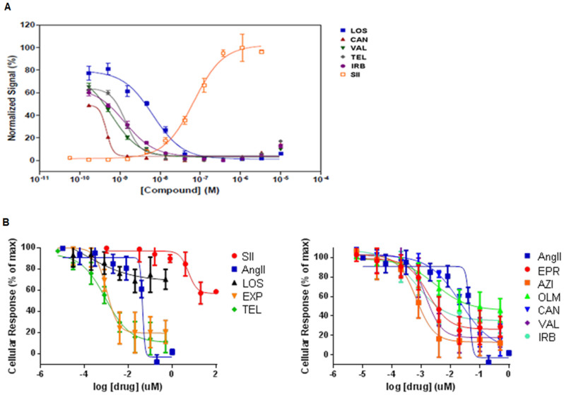
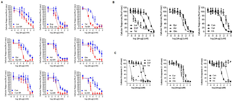
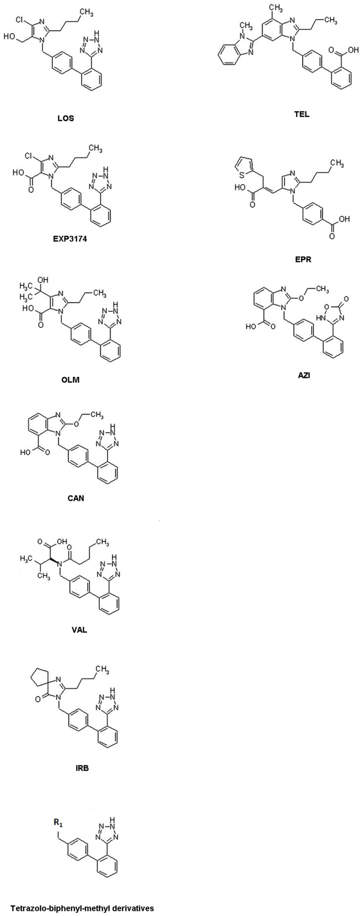
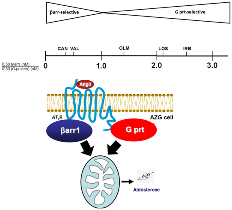
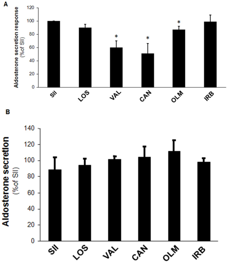
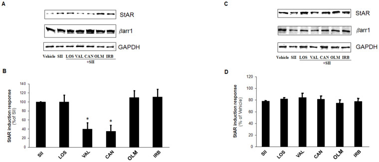
The known angiotensin II (AngII) physiological effect of aldosterone synthesis and secretion is mediated by either Gq/11 proteins or βarrestin1 (βarr1), both of which can couple to its type 1 receptors (AT₁Rs), present in adrenocortical zona glomerulosa (AZG) cell membranes. In the present study, we examined the relative potencies of all the currently used in the clinic AT₁R antagonist drugs (angiotensin receptor blockers, ARBs, or sartans) at preventing activation of these two signaling mediators (G proteins and βarrs) at the AngII-bound AT1R and, consequently, at suppression of aldosterone in vitro. All ARBs were found to be potent inhibitors of G protein activation at the AT₁R. However, candesartan and valsartan were the most potent at blocking AngII-induced βarr activation at this receptor, among the tetrazolo-biphenyl-methyl derivatives, translating into excellent efficacies at aldosterone suppression in H295R cells. Conversely, irbesartan and losartan were largely G protein-selective inhibitors at the AT₁R, with very low potency towards βarr inhibition. As a result, they were very weak suppressors of βarr1-dependent aldosterone production in H295R cells. These findings provide important pharmacological insights into the drug class of ARBs and medicinal chemistry insights for future drug development in the field of AngII antagonism.


Figures

Figure 1
Figure 1. Lack of agonist activity of ARBs for AT1R-induced G protein or βarr activation.
(A) Dose-response curves of each drug for βarr activation at the AT1R, based on the DiscoveRx PathHunterTM β-Arrestin assay system. (B) Dose-response curves of each drug for acute G protein or βarr activation derived from the CellKey assay. LOS: Losartan; EXP: EXP3174; TEL: Telmisartan; EPR: Eprosartan; AZI: Azilsartan; OLM: Olmesartan; CAN: Candesartan; VAL: Valsartan; IRB: Irbesartan.
Figure 2
Figure 2. In vitro potency of sartans at blocking AT1R-induced G protein or βarr activation.
(A) Dose-response curves for inhibition of AngII-induced AT1R-βarr binding or for inhibition of AngII-induced AT1R-G protein coupling (early response, ER) for each compound, as derived from the CellKey assay. (B) Dose-response curves of each drug for βarr activation at the AngII-bound AT1R, based on the DiscoveRx PathHunterTM β-Arrestin assay system. (C) AngII-induced G protein inhibition potencies based on the FLIPR calcium assay (Molecular Devices). Los: Losartan; Exp: EXP3174; Tel: Telmisartan; Epr: Eprosartan; Azi: Azilsartan; Olm: Olmesartan; Can: Candesartan; Val: Valsartan; Irb: Irbesartan.
Figure 3
Figure 3. Chemical structures of the drugs investigated in the present study.
LOS: Losartan; EXP: EXP3174; TEL: Telmisartan; EPR: Eprosartan; AZI: Azilsartan; OLM: Olmesartan; CAN: Candesartan; VAL: Valsartan; IRB: Irbesartan; R1: Any alkyl- or aryl-substitution.
Figure 4
Figure 4. Ranking of “selectivity” for G protein (G prt) or βarr inhibition at the AT1R and association with AT1R-dependent aldosterone suppression.
Ratios <1.0 denote relative selectivity for βarr inhibition (the lower the ratio, the higher the selectivity), ratios >1.0 denote relative selectivity for G protein inhibition (and the higher the ratio, the higher the selectivity), whereas a ratio of 1.0 denotes no selectivity. LOS: losartan; CAN: candesartan; VAL: valsartan; IRB: irbesartan; OLM: olmesartan.
Figure 5
Figure 5. Efficacy of ARBs at suppressing in vitro aldosterone secretion from H295R cells.
Aldosterone secretion in vitro by H295R cells stimulated for 6 hrs with 10 μM SII alone (SII) or in the presence of 10 μM of each of the sartans tested (losartan-LOS, valsartan-VAL, candesartan-CAN, olmesartan-OLM, or irbesartan-IRB). (A) Data from cells transfected to overexpress βarr1. Data are shown as % of the response to SII alone. *, p < 0.05, vs. SII, n = 4 independent experiments/treatment. (B) Data from cells transfected to overexpress the dominant negative V53D βarr1 mutant (23). Data are shown as % of the response to SII alone. No significant differences were observed among treatments, nor did SII induce any response, as expected, since endogenous βarr1 is blocked. n = 3 independent experiments/treatment.
Figure 6
Figure 6. Efficacy of ARBs at preventing StAR upregulation in SII-stimulated H295R cells.
(A,B) Western blotting for StAR protein levels in H295R cells transfected to overexpress βarr1 and treated for 6 hrs with 10 μM SII alone (SII) or in the presence of 10 μM of each of the sartans tested. Representative blots of 3 independent experiments are shown in (A), including blots for βarr1 to confirm its overexpression and for GAPDH (glyceraldehyde 3-phosphate dehydrogenase) as loading control, and the StAR protein induction (as % of the SII response), as derived by densitometric quantification, is shown in (B). *, p < 0.05, n = 3 independent experiments/treatment. Blots shown have been cropped to fit space requirements and run under the same experimental conditions (same gel) (the full length blots are shown in Supplementary Information). (C,D) Western blotting for StAR protein levels in dominant negative βarr1 mutant-transfected H295R cells and treated as in (A–B). Representative blots are shown in (C), including blots for the dominant negative βarr1 mutant to confirm its overexpression and for GAPDH as loading control, and the StAR protein induction (as % of vehicle-no stimulation), as derived by densitometric quantification, is shown in (D). No significant differences were observed among treatments, nor did any treatment cause any induction in StAR levels. Blots shown have been cropped to fit space requirements and run under the same experimental conditions (same gel) (the full length blots are shown in Supplementary Information). n = 3 independent experiments/treatment. LOS: Losartan-; VAL: Valsartan; CAN: Candesartan; OLM: Olmesartan; IRB: Irbesartan.
Figure 7
Figure 7. In vivo effects of candesartan and valsartan in post-MI rats overexpressing adrenal βarr1.
(A) Trichrome-Masson's staining in myocardial cross-sections from post-MI rats overexpressing βarr1 specifically in their adrenals and treated for 7 days with either vehicle or 10 mg/kg body weight/day candesartan or 40 mg/kg body weight/day irbesartan (all via drinking water). Blue denotes collagen fibers, red denotes muscle fibers, and black represents cell nuclei. Representative images are shown from 5–6 rat hearts stained per group, along with staining in sham-operated rat hearts, in which no blue staining was detectable. (B,C) Heart mRNA levels of (B) collagen I (Col1a1) and brain natriuretic peptide (BNP) (C) in these rats at the end of the 7-day drug treatments. Values for post-MI rats receiving AdGFP in their adrenals instead of Adβarr1 (i.e. not overexpressing βarr1 in their adrenals) were used as reference. *, p < 0.05, vs. Candesartan (Adβarr1), n = 5 rat hearts/group.

References

    1. Weber K. T. Aldosterone in congestive heart failure. N. Engl. J. Med. 345, 1689–1697 (2001). - PubMed
    1. Rainey W. E., Saner K. & Schimmer B. P. Adrenocortical cell lines. Mol. Cell. Endocrinol. 228, 23–38 (2004). - PubMed
    1. Luttrell L. M. & Gesty-Palmer D. Beyond desensitization: physiological relevance of arrestin-dependent signaling. Pharmacol. Rev. 62, 305–330 (2010). - PMC - PubMed
    1. Lymperopoulos A. et al. An adrenal beta-arrestin 1-mediated signaling pathway underlies angiotensin II-induced aldosterone production in vitro and in vivo. Proc. Natl. Acad. Sci. USA. 106, 5825–5830 (2009). - PMC - PubMed
    1. Lymperopoulos A. et al. Adrenal beta-arrestin 1 inhibition in vivo attenuates post-myocardial infarction progression to heart failure and adverse remodeling via reduction of circulating aldosterone levels. J. Am. Coll. Cardiol. 57, 356–365 (2011). - PMC - PubMed

Publication types

MeSH terms