Case Reports
doi: 10.11622/smedj.2014168.
An unusual presentation of primary malignant B-cell-type dural lymphoma
Affiliations
- PMID: 25631982
- PMCID: PMC4294017
- DOI: 10.11622/smedj.2014168
Item in Clipboard
Case Reports
An unusual presentation of primary malignant B-cell-type dural lymphoma
Singapore Med J.
2014 Nov.
Abstract
Primary malignant B-cell-type dural lymphoma is a rare subtype of primary central nervous system lymphoma (PCNSL). We herein report an unusual case of diffuse B-cell lymphoma that presents as a chronic subdural haematoma without extracranial involvement. The notable aspects of this case include the patient's immunocompetence, a short clinical history of symptom onset, rapid neurological deterioration and a fi nal diagnosis of high-grade PCNSL. This case highlights the challenges neurosurgeons face, especially in the emergency setting, when the disease manifests in varied presentations.
Figures

Pre-contrast axial CT image shows the patient’s brain.
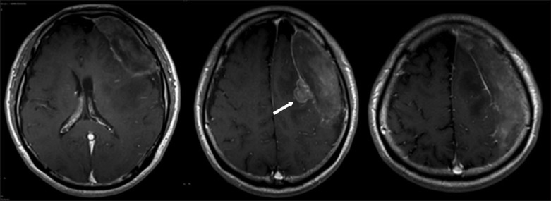
T1-weighted post-gadolinium MR image shows the extra-axial polypoidal nipple, which was thought to be a proliferation of granulation tissue (arrow).

Photomicrographs of the lesional tissue show (a) the dura at the bottom right, under low magnification (Haematoxylin & eosin); and (b) sheets of neoplastic cells with brisk mitotic activity, under high magnification (Haematoxylin & eosin). The neoplastic cells contain enlarged vesicular nuclei with small but conspicuous nucleoli, and show (c) strong and diffuse immunostaining for CD20; and (d) marked MIB-1 labelling index.

Photomicrographs of the immunostains of the neoplastic cells show positive tests for (a) BCL2 and (b) CD43. Photomicrographs of the scattered cells show (c) positive staining for CycD1; and (d) a small population of reactive CD5-positive T lymphocytes.
References
-
- Chen TC, Abrey LE. Primary central nervous system lymphomas. J Neurosurg. 2006;21:1. - PubMed
-
- Schabet M. Epidemiology of primary CNS lymphoma. J Neurooncol. 1999;43:199–201. - PubMed
-
- Sacho RH, Kogels M, du Plessis D, Jowitt S, Josan VA. Primary diffuse large B-cell nervous system lymphoma presenting as an acute space-occupying subdural mass. J Neurosurg. 2010;113:384–7. - PubMed
-
- Beriwal S, Hou JS, Miyamoto C, Garcia-Young JA. Primary dural low grade BCL-2 negative follicular lymphoma: a case report. J Neurooncol. 2003;61:23–5. - PubMed
Publication types
MeSH terms
LinkOut - more resources
Full Text Sources
Other Literature Sources
Medical