. 2015 Mar 7;282(1802):20142857.
doi: 10.1098/rspb.2014.2857.

A 'NanoSuit' surface shield successfully protects organisms in high vacuum: observations on living organisms in an FE-SEM

Affiliations

A 'NanoSuit' surface shield successfully protects organisms in high vacuum: observations on living organisms in an FE-SEM

Yasuharu Takaku et al. Proc Biol Sci. .

Abstract

Although extremely useful for a wide range of investigations, the field emission scanning electron microscope (FE-SEM) has not allowed researchers to observe living organisms. However, we have recently reported that a simple surface modification consisting of a thin extra layer, termed 'NanoSuit', can keep organisms alive in the high vacuum (10(-5) to 10(-7) Pa) of the SEM. This paper further explores the protective properties of the NanoSuit surface-shield. We found that a NanoSuit formed with the optimum concentration of Tween 20 faithfully preserves the integrity of an organism's surface without interfering with SEM imaging. We also found that electrostatic charging was absent as long as the organisms were alive, even if they had not been coated with electrically conducting materials. This result suggests that living organisms possess their own electrical conductors and/or rely on certain properties of the surface to inhibit charging. The NanoSuit seems to prolong the charge-free condition and increase survival time under vacuum. These findings should encourage the development of more sophisticated observation methods for studying living organisms in an FE-SEM.

Keywords: NanoSuit; field emission scanning electron microscope; high vacuum; living organism; surface shield effect.


Figures

Figure 1.
Figure 1.
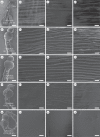
Comparison of mosquito larvae treated with different concentrations of Tween 20 (TW20) and observed for 30 min in the SEM. The small white squares in a, e, i, m, q indicate the position of images shown at high magnification in bd, fh, jl, np, rt. Each image comes from a different treated larva. The thin TW20 NanoSuit in the specimens shown in ad and h was not sufficient to protect them from dehydration and/or resulted in electrostatic charging (b,c,d,h). The thick TW20 NanoSuit (m,q) prevented imaging of the surface fine structure (np, rt). Scale bars, 500 μm (a,e,i,m,q), 500 nm (bd, fh, jl, np, rt).
Figure 2.
Figure 2.
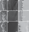
Comparison of SEM and TEM images of mosquito larvae treated with 1% TW20 solution (ac), with an additional coating of gold (df) and with 50% TW20 solution (gi). SEM images (a,b,d,e,g,h) and TEM images (c,f,i). The white squares in a, d, g indicate the position of images magnified in b, e, h. The layer between the arrowheads in c, f, i indicates the newly formed NanoSuit. Scale bars, 500 μm (a,d,g), 500 nm (b,e,h), 300 nm (c,f,i).
Figure 3.
Figure 3.
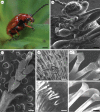
Observations of living insects by light and electron microscopy. (a) Stereo dissecting microscopic observation of the leaf beetle Lilioceris merdigera. (b) SEM observation of the living beetle. (cg) Direct SEM observations of the ‘hairy’ adhesive organ. (c) Tip of the left foreleg's tarsus, showing five independent tarsomeres; first (1) to fifth (5) tarsomeres, respectively, from the proximal end. (d,e) Observations at higher magnifications of the hairy pads. (f,g) Electrostatic charge quickly appeared on dead specimens even if pre-treated with TW20 solution. Scale bars, 500 μm (a), 200 μm (b), 100 μm (c), 10 μm (d,f), 2 μm (e,g).
Figure 4.
Figure 4.
Electron beam-induced deposits appeared in the observation field, usually as a square shape, in a conventional SEM preparation (top). The surface of a living animal protected with a NanoSuit showed less deposit (bottom). The white squares in a and c are shown at high magnifications in b and d, respectively. Scale bars, 200 μm.

References

    1. Knoll M. 1935. Aufladepotentiel und Sekundäremission elektronenbestrahlter Körper. Z. Techn. Phys. 16, 467–475.
    1. Small EB, Marszalek DS. 1969. Scanning electron microscopy of fixed, frozen, and dried protozoa. Science 163, 1064–1065. (10.1126/science.163.3871.1064) - DOI - PubMed
    1. Suzuki E. 2002. High-resolution scanning electron microscopy of immunogold-labelled cells by the use of thin plasma coating of osmium. J. Microsc. 208, 153–157. (10.1046/j.1365-2818.2002.01082.x) - DOI - PubMed
    1. Symondson WOC, Williams IB. 2003. Low-vacuum electron microscopy of carabid chemoreceptors: a new tool for the identification of live and valuable museum specimens. Entomol. Exp. Appl. 85, 75–82. (10.1046/j.1570-7458.1997.00235.x) - DOI
    1. Danilatos GD. 1991. Review and outline of environmental SEM at present. J. Microsc. 162, 391–402. (10.1111/j.1365-2818.1991.tb03149.x) - DOI

LinkOut - more resources