Voluntary suppression of hyperthermia-induced hyperventilation mitigates the reduction in cerebral blood flow velocity during exercise in the heat
- PMID: 25632021
- PMCID: PMC4398858
- DOI: 10.1152/ajpregu.00419.2014
Voluntary suppression of hyperthermia-induced hyperventilation mitigates the reduction in cerebral blood flow velocity during exercise in the heat
Abstract
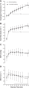
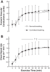
Hyperthermia during prolonged exercise leads to hyperventilation, which can reduce arterial CO2 pressure (PaCO2 ) and, in turn, cerebral blood flow (CBF) and thermoregulatory response. We investigated 1) whether humans can voluntarily suppress hyperthermic hyperventilation during prolonged exercise and 2) the effects of voluntary breathing control on PaCO2 , CBF, sweating, and skin blood flow. Twelve male subjects performed two exercise trials at 50% of peak oxygen uptake in the heat (37°C, 50% relative humidity) for up to 60 min. Throughout the exercise, subjects breathed normally (normal-breathing trial) or they tried to control their minute ventilation (respiratory frequency was timed with a metronome, and target tidal volumes were displayed on a monitor) to the level reached after 5 min of exercise (controlled-breathing trial). Plotting ventilatory and cerebrovascular responses against esophageal temperature (Tes) showed that minute ventilation increased linearly with rising Tes during normal breathing, whereas controlled breathing attenuated the increased ventilation (increase in minute ventilation from the onset of controlled breathing: 7.4 vs. 1.6 l/min at +1.1°C Tes; P < 0.001). Normal breathing led to decreases in estimated PaCO2 and middle cerebral artery blood flow velocity (MCAV) with rising Tes, but controlled breathing attenuated those reductions (estimated PaCO2 -3.4 vs. -0.8 mmHg; MCAV -10.4 vs. -3.9 cm/s at +1.1°C Tes; P = 0.002 and 0.011, respectively). Controlled breathing had no significant effect on chest sweating or forearm vascular conductance (P = 0.67 and 0.91, respectively). Our results indicate that humans can voluntarily suppress hyperthermic hyperventilation during prolonged exercise, and this suppression mitigates changes in PaCO2 and CBF.
Keywords: cerebral blood flow; hyperpnea; hyperthermia; voluntary control of breathing.
Copyright © 2015 the American Physiological Society.
Figures


Similar articles
-
Respiratory mechanics and cerebral blood flow during heat-induced hyperventilation and its voluntary suppression in passively heated humans.Physiol Rep. 2019 Jan;7(1):e13967. doi: 10.14814/phy2.13967. Physiol Rep. 2019. PMID: 30637992 Free PMC article.
-
Effect of hypocapnia on the sensitivity of hyperthermic hyperventilation and the cerebrovascular response in resting heated humans.J Appl Physiol (1985). 2018 Jan 1;124(1):225-233. doi: 10.1152/japplphysiol.00232.2017. Epub 2017 Sep 28. J Appl Physiol (1985). 2018. PMID: 28970199
-
Effect of short-term exercise-heat acclimation on ventilatory and cerebral blood flow responses to passive heating at rest in humans.J Appl Physiol (1985). 2015 Sep 1;119(5):435-44. doi: 10.1152/japplphysiol.01049.2014. Epub 2015 Jul 9. J Appl Physiol (1985). 2015. PMID: 26159763
-
Components and mechanisms of thermal hyperpnea.J Appl Physiol (1985). 2006 Aug;101(2):655-63. doi: 10.1152/japplphysiol.00210.2006. Epub 2006 Mar 24. J Appl Physiol (1985). 2006. PMID: 16565352 Review.
-
Cerebral oxygenation and hyperthermia.Front Physiol. 2014 Mar 4;5:92. doi: 10.3389/fphys.2014.00092. eCollection 2014. Front Physiol. 2014. PMID: 24624095 Free PMC article. Review.
Cited by
-
Middle cerebral artery blood flow velocity during a 4 km cycling time trial.Eur J Appl Physiol. 2017 Jun;117(6):1241-1248. doi: 10.1007/s00421-017-3612-2. Epub 2017 Apr 13. Eur J Appl Physiol. 2017. PMID: 28409398 Clinical Trial.
-
Characteristics of hyperthermia-induced hyperventilation in humans.Temperature (Austin). 2016 Feb 18;3(1):146-60. doi: 10.1080/23328940.2016.1143760. eCollection 2016 Jan-Mar. Temperature (Austin). 2016. PMID: 27227102 Free PMC article. Review.
-
Respiratory mechanics and cerebral blood flow during heat-induced hyperventilation and its voluntary suppression in passively heated humans.Physiol Rep. 2019 Jan;7(1):e13967. doi: 10.14814/phy2.13967. Physiol Rep. 2019. PMID: 30637992 Free PMC article.
-
Physiological Function during Exercise and Environmental Stress in Humans-An Integrative View of Body Systems and Homeostasis.Cells. 2022 Jan 24;11(3):383. doi: 10.3390/cells11030383. Cells. 2022. PMID: 35159193 Free PMC article. Review.
-
Intradermal administration of ATP augments methacholine-induced cutaneous vasodilation but not sweating in young males and females.Am J Physiol Regul Integr Comp Physiol. 2015 Oct 15;309(8):R912-9. doi: 10.1152/ajpregu.00261.2015. Epub 2015 Aug 19. Am J Physiol Regul Integr Comp Physiol. 2015. PMID: 26290105 Free PMC article.
References
-
- Albert RE. Sweat suppression by forced breathing in man. J Appl Physiol 20: 134–136, 1965. - PubMed
-
- Asmussen E, Johansen SH, Jorgensen M, Nielsen M. On the nervous factors controlling respiration and circulation during exercise experiments with curarization. Acta Physiol Scand 63: 343–350, 1965. - PubMed
-
- Bain AR, Smith KJ, Lewis NC, Foster GE, Wildfong KW, Willie CK, Hartley GL, Cheung SS, Ainslie PN. Regional changes in brain blood flow during severe passive hyperthermia: effects of PaCO2 and extracranial blood flow. J Appl Physiol 115: 653–659, 2013. - PubMed
-
- Beaudin AE, Clegg ME, Walsh ML, White MD. Adaptation of exercise ventilation during an actively-induced hyperthermia following passive heat acclimation. Am J Physiol Regul Integr Comp Physiol 297: R605–R614, 2009. - PubMed
Publication types
MeSH terms
LinkOut - more resources
Full Text Sources
Other Literature Sources
Medical

