. 2015 Feb 24;131(8):695-708.
doi: 10.1161/CIRCULATIONAHA.114.013708. Epub 2015 Jan 28.

Dysfunction in the βII spectrin-dependent cytoskeleton underlies human arrhythmia

Affiliations

Dysfunction in the βII spectrin-dependent cytoskeleton underlies human arrhythmia

Sakima A Smith et al. Circulation. .

Abstract

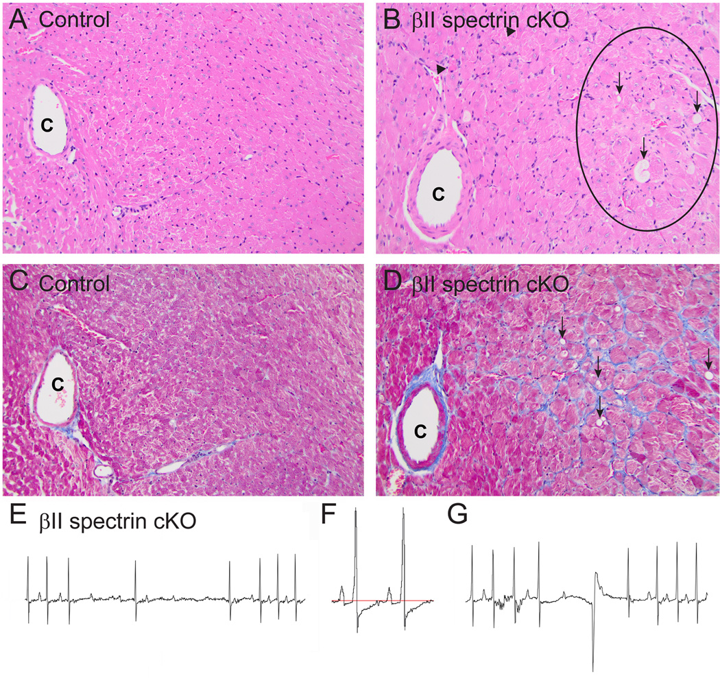
Background: The cardiac cytoskeleton plays key roles in maintaining myocyte structural integrity in health and disease. In fact, human mutations in cardiac cytoskeletal elements are tightly linked to cardiac pathologies, including myopathies, aortopathies, and dystrophies. Conversely, the link between cytoskeletal protein dysfunction and cardiac electric activity is not well understood and often overlooked in the cardiac arrhythmia field.

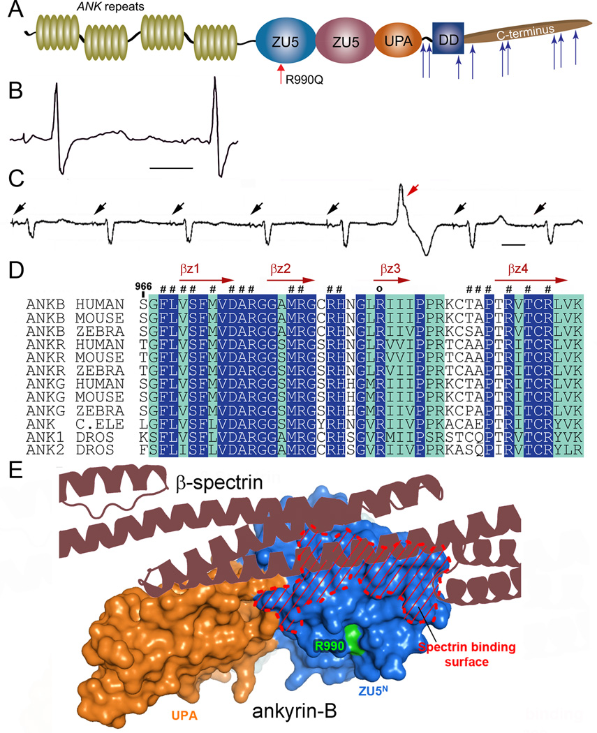
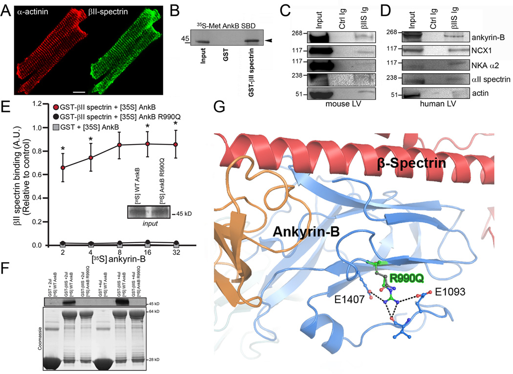
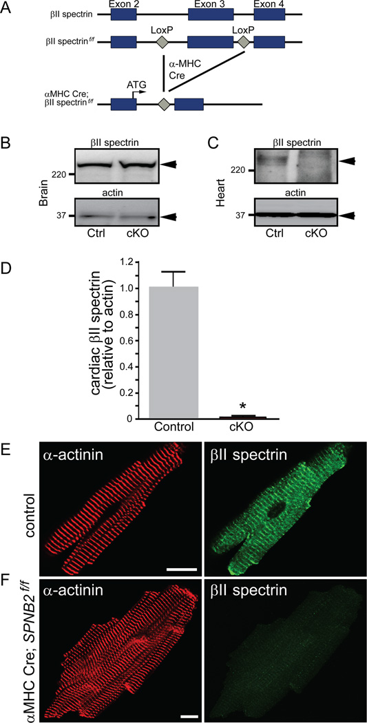
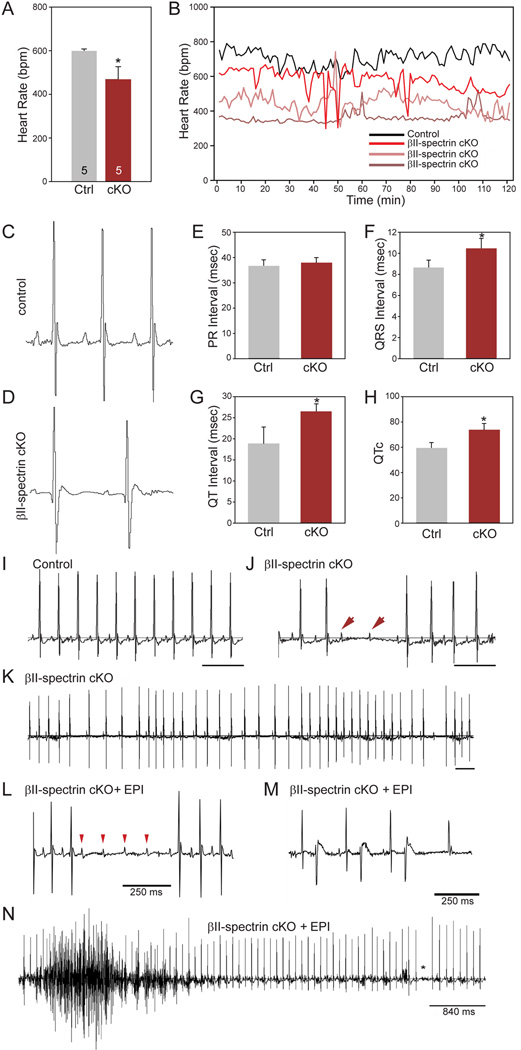
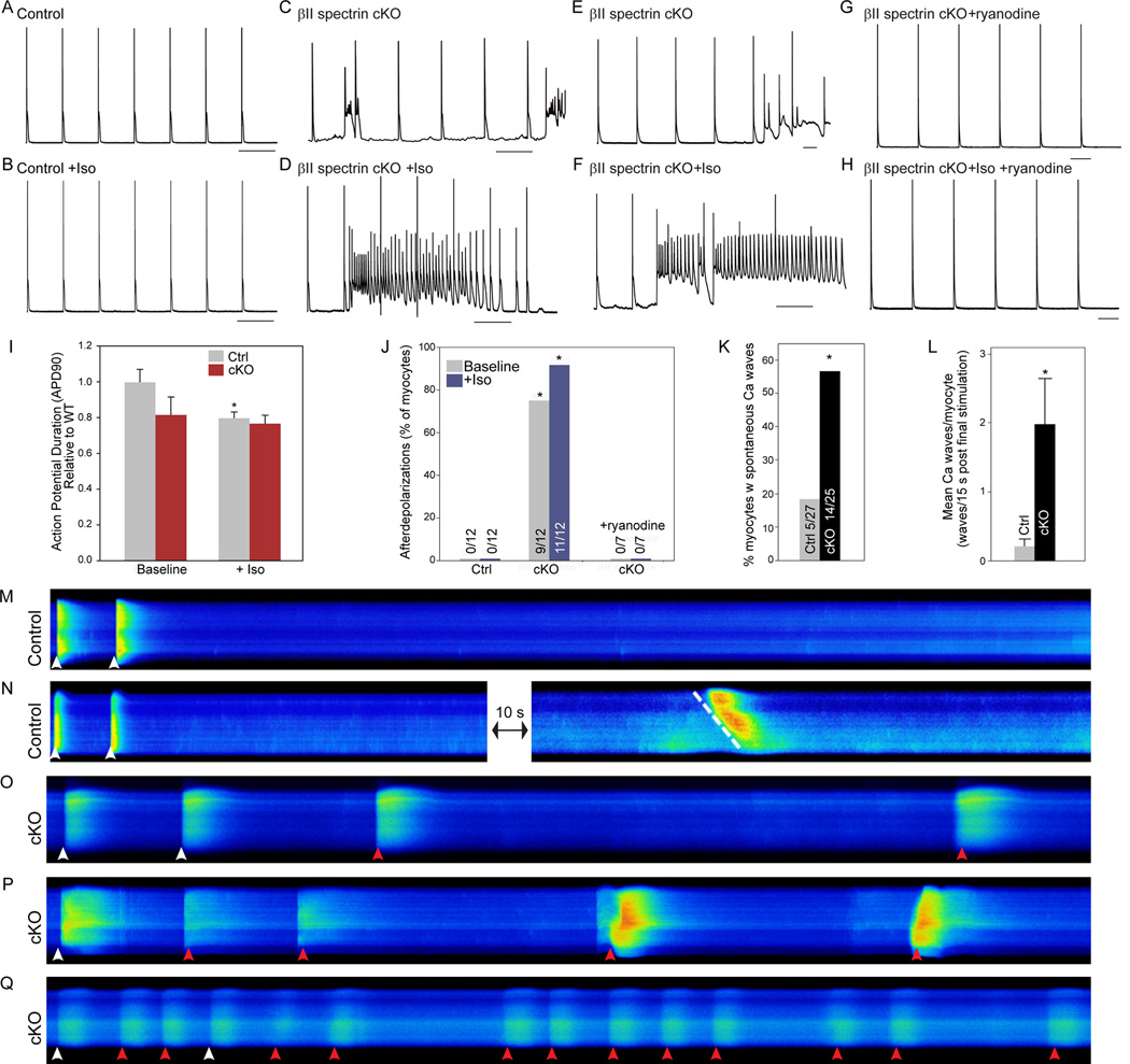
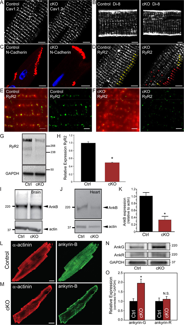
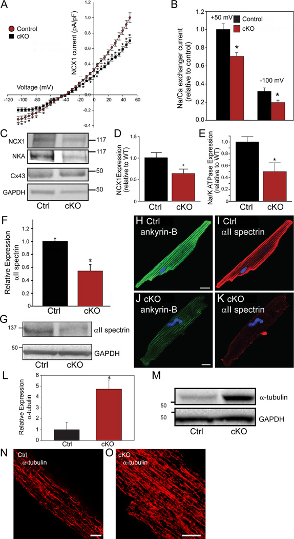
Methods and results: Here, we uncover a new mechanism for the regulation of cardiac membrane excitability. We report that βII spectrin, an actin-associated molecule, is essential for the posttranslational targeting and localization of critical membrane proteins in heart. βII spectrin recruits ankyrin-B to the cardiac dyad, and a novel human mutation in the ankyrin-B gene disrupts the ankyrin-B/βII spectrin interaction, leading to severe human arrhythmia phenotypes. Mice lacking cardiac βII spectrin display lethal arrhythmias, aberrant electric and calcium handling phenotypes, and abnormal expression/localization of cardiac membrane proteins. Mechanistically, βII spectrin regulates the localization of cytoskeletal and plasma membrane/sarcoplasmic reticulum protein complexes, including the Na/Ca exchanger, ryanodine receptor 2, ankyrin-B, actin, and αII spectrin. Finally, we observe accelerated heart failure phenotypes in βII spectrin-deficient mice.

Conclusions: Our findings identify βII spectrin as critical for normal myocyte electric activity, link this molecule to human disease, and provide new insight into the mechanisms underlying cardiac myocyte biology.

Keywords: arrhythmias, cardiac; catecholaminergic polymorphic ventricular arrhythmia; cytoskeleton; ion channels; protein transport; ventricular tachycardia.


Figures

Figure 1
Figure 1
Ankyrin-B arrhythmia variant identified in conserved spectrin-binding domain. (A) Ankyrin-B includes ANK repeats, a spectrin-binding domain comprised of two ZU5 and one UPA domain, and a regulatory domain comprised of a death and C-terminus. Identified ANK2 loss-of-function mutations are noted by blue arrows and novel p.R990Q variant is indicated in red. (B) ANK2 p.R990Q proband displays QT-prolongation. (C) 10 second rhythm strip in p.R990Q proband demonstrating atrial demand pacing (black arrows) with premature ventricular contraction (red arrow). (D) Sequence alignment of ankyrin-B spectrin-binding sequence. Residues that are absolutely conserved and highly conserved are in blue and green, respectively. Secondary structural elements are indicated above the alignment. p.R990Q is conserved across species and marked with “o”. (E) Structure of the ZU5N-UPA tandem of ankyrin-B SBD reveals the spectrin-binding surface and location of p.R990 (green).
Figure 2
Figure 2
Ankyrin-B/βII spectrin complex is blocked by human disease mutation. (A) βII spectrin (green) is localized in a striated pattern in isolated mouse myocytes (co-labelled with α-actinin, red). Scale=10 μm. (B) Radiolabelled ankyrin-B (spectrin-binding domain) associates with GST βII spectrin fusion protein, but not GST. (C-D) βII spectrin (βIIS) Ig co-immunoprecipitates ankyrin-B, NCX1, Na/K ATPase, αII spectrin, and actin from detergent soluble lysates of non-failing mouse and human LV. (E) Human ANK2 p.R990Q arrhythmia variant displays aberrant βII spectrin-binding. Data in inset represents equal inputs for experiments. Curves denote binding for GST-βII spectrin or GST alone with a concentration range of radiolabelled ankyrin-B or radiolabelled ankyrin-B R990Q (n=5/group; * represents p<0.05). Panel F represents primary binding data and Coomassie Blue-stained gels for fusion proteins in E. (G) Ribbon structure from co-crystal structure of ankyrin-B/βII spectrin. Human p.R990Q variant shown in green.
Figure 3
Figure 3
Generation and validation of mice lacking βII spectrin in cardiomyocytes. (A) Targeting strategy to generate Cre-dependent loss of cardiac βII spectrin. βII spectrin in brain (B) and heart (C) of control and βII spectrin cKO mice. (D) βII spectrin levels in control (n=5) versus βII spectrin cKO hearts (n=5; p<0.05). (E) βII spectrin localization in control and βII spectrin cKO adult ventricular myocytes. Bar= 10 μm.
Figure 4
Figure 4
Loss of βII spectrin causes bradycardia, rate variability, and arrhythmia. (A-B) βII spectrin cKO mice display reduced heart rate (n=5) as assessed by telemetry compared to control mice (n=5, p<0.05). (B) Heart rates of control and three βII spectrin cKO mice that show bradycardia and rate variability. (C-D) ECGs from control and βII spectrin cKO mouse showing increased RR, QRS, and QT intervals. Mean data for parameters are shown inE-H (n=5 mice/genotype; p<0.05). (I-J) Control ECG recording over 1-second demonstrating no R-R variability or heart block vs. ECG recording from βII spectrin cKO littermate demonstrating type II heart block, confirmed by P-waves (arrow heads) without ventricular conduction. (K) 3-second ECG recording of βII spectrin cKO mouse demonstrating significant R-R variability with heart block. (L-N) βII spectrin cKO mice demonstrate severe arrhythmia phenotypes and death following injection of epinephrine. Examples include (L) four sinus P-waves (arrow heads) without ventricular conduction consistent with type II AV block, (M) bigeminy, and (N) polymorphic ventricular arrhythmia.
Figure 5
Figure 5
βII spectrin cKO myocytes display electrical instability, afterdepolarizations, and aberrant Ca waves. Action potential measurements of (A-B) control and (C-D) βII spectrin cKO myocytes measured at baseline at 0.5 Hz pacing protocol ± 1μM Iso. (E-F) Electrical instability was present in βII spectrin cKO myocytes independent of pacing frequency (shown at 1 Hz). (G-H) Ryanodine (100 nM) blocked abnormal electrical instability of βII spectrin cKO myocytes ± Iso. (I) Mean APD90 of control and βII spectrin cKO myocytes ±Iso (n> 10 myocytes genotype; p<0.05 for control vs. control + Iso). (J) Prevalence of afterdepolarizations for control and βII spectrin cKO myocytes ±Iso and in the presence of ryanodine (n>10 myocytes/treatment; p<0.05 for control versus cKO at baseline; p<0.05 for control +Iso versus cKO +Iso). (K-L) βII spectrin cKO myocytes were more likely to form spontaneous Ca2+ waves (n=27 control, n=25 βII spectrin cKO; p<0.05). (M-Q) Linescan images of fluo-4-loaded myocytes field stimulated at 0.5 Hz. Following stimulation, myocytes were continuously monitored for spontaneous Ca2+ wave formation for 15 sec. (M-N) Control myocytes with no spontaneous wave activity. When waves formed they were slow moving (N, dashed white line). (O-Q) Spontaneous waves in βII spectrin cKO myocytes (red arrowheads). Stimulated transients are indicated by white arrowheads.
Figure 6
Figure 6
βII spectrin is required for organization of calcium release units and ankyrin-B. Like control mouse myocytes, βII spectrin cKO myocytes display normal localization of transverse-tubule Cav1.2 (A), transverse-tubule organization (B, visualized by Di-8-ANEPPs), and intercalated disc N-cadherin (C). In contrast, RyR2 expression was reduced and heterogeneous in βII spectrin cKO myocytes (D, red *). (E-F) RyR2 expression was further analyzed by total internal reflection fluorescence (TIRF) and super-resolution imaging. (E-F, left) Overlay of a TIRF image (red) and the corresponding super-resolution image (green) of RyR2 in control (E) and βII spectrin cKO (F) cardiomyocyte. (E-F, right) Super-resolution images of RyR2 in control and βII spectrin cKO myocytes. Note that data in F was collected from area of RyR2 heterogeneity (red * cKO panel D, right) that illustrates reduced RyR2 cluster size and intensity. Scale bar for E-F: 1000 nm. (G-H) RyR2 levels were significantly reduced in βII spectrin cKO myocytes (n=4 hearts/genotype, p<0.05). (I-K) Ankyrin-B levels are decreased in the heart but not brain of βII spectrin cKO mice (n=5 hearts/genotype; p<0.05). (L-M) Ankyrin-B shows reduced expression and abnormal targeting in βII spectrin cKO versus control myocytes (Bar=10 microns). (N-O) Ankyrin-G, but not ankyrin-R levels are decreased in the heart of βII spectrin cKO mice (n=5) versus control mice (n=6; p<0.05).
Figure 7
Figure 7
βII spectrin deficiency results in reduced expression of ankyrin-B-associated membrane proteins and abnormal myocyte cytoskeletal organization. (A-B) βII spectrin cKO myocytes (n=12) display reduced INCX compared with control mouse myocytes (n=14; p<0.05). (C-E) βII spectrin cKO hearts display reduced NCX and Na/K ATPase expression but normal expression of Cx43 compared with control hearts (n=4 hearts/genotype; p<0.05). (F-K) βII spectrin cKO hearts (n=5) display reduced αII spectrin expression and localization compared with control hearts (n=5) by immunoblot and immunostaining (p<0.05). (L-O) βII spectrin cKO hearts display significant increase in α-tubulin expression by immunoblot and immunostaining compared to control hearts (n=5 hearts/genotype for panel L; p<0.05). Bar=10 μM for panels N-O.
Figure 8
Figure 8
βII spectrin cKO mice show severe damage and electrical phenotypes following aortic banding. (A-B) Unlike control TAC mice, βII spectrin cKO mice displayed widespread myocardial degeneration of the LV free wall and septum, characterized by vacuolation (arrows), pallor (inside circle), and necrosis of myocytes (C=coronary artery; H&E staining; 200x). (C-D) βII spectrin cKO hearts further displayed increased interstitial fibrosis (blue) of connective tissue compared with control sections (vacuolation noted by arrows; 200x). Electrical phenotypes observed in βII spectrin cKO and not control mice included (E) AV conduction defects, (F) ST segment depression, and (G) intermittent PVCs and junctional beats.

Comment in

  • Cytoskeleton regulation of ion channels.
    Fu Y, Xiao S, Hong T, Shaw RM. Fu Y, et al. Circulation. 2015 Feb 24;131(8):689-91. doi: 10.1161/CIRCULATIONAHA.115.015216. Epub 2015 Jan 28. Circulation. 2015. PMID: 25632042 Free PMC article. No abstract available.

References

    1. Moulik M, Vatta M, Witt SH, Arola AM, Murphy RT, McKenna WJ, Boriek AM, Oka K, Labeit S, Bowles NE, Arimura T, Kimura A, Towbin JA. Ankrd1, the gene encoding cardiac ankyrin repeat protein, is a novel dilated cardiomyopathy gene. J Am Coll Cardiol. 2009;54:325–333. - PMC - PubMed
    1. Lammerding J, Schulze PC, Takahashi T, Kozlov S, Sullivan T, Kamm RD, Stewart CL, Lee RT. Lamin a/c deficiency causes defective nuclear mechanics and mechanotransduction. J Clin Invest. 2004;113:370–378. - PMC - PubMed
    1. van Tintelen JP, Entius MM, Bhuiyan ZA, Jongbloed R, Wiesfeld AC, Wilde AA, van der Smagt J, Boven LG, Mannens MM, van Langen IM, Hofstra RM, Otterspoor LC, Doevendans PA, Rodriguez LM, van Gelder IC, Hauer RN. Plakophilin-2 mutations are the major determinant of familial arrhythmogenic right ventricular dysplasia/cardiomyopathy. Circulation. 2006;113:1650–1658. - PubMed
    1. Stroud MJ, Banerjee I, Veevers J, Chen J. Linker of nucleoskeleton and cytoskeleton complex proteins in cardiac structure, function, and disease. Circ Res. 2014;114:538–548. - PMC - PubMed
    1. Schwartz PJ, Ackerman MJ, George AL, Jr, Wilde AA. Impact of genetics on the clinical management of channelopathies. J Am Coll Cardiol. 2013;62:169–180. - PMC - PubMed

Publication types

MeSH terms