

Quantification of sulfatides and lysosulfatides in tissues and body fluids by liquid chromatography-tandem mass spectrometry
- PMID: 25632048
- PMCID: PMC4373750
- DOI: 10.1194/jlr.M057232
Quantification of sulfatides and lysosulfatides in tissues and body fluids by liquid chromatography-tandem mass spectrometry
Abstract
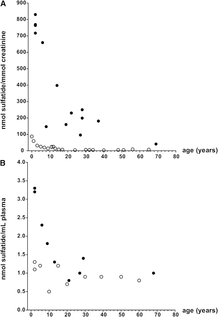
Sulfatides are found in brain as components of myelin, oligodendrocytes, and neurons but are also present in various visceral tissues. Metachromatic leukodystrophy (MLD) is an inherited lysosomal storage disorder caused by a deficiency of arylsulfatase A, leading to severe white matter disease due to the accumulation of sulfatides and lysosulfatides. To study the physiological role of sulfatides, accessible and sensitive quantitative methods are required. We developed a sensitive LC/MS/MS method to quantify total sulfatide and lysosulfatide content as well as individual molecular species in urine and plasma from MLD patients and plasma and tissues from an MLD mouse model. Our results demonstrate that the method can quantify a wide range of sulfatide concentrations and can be used to quantify total sulfatide content and levels of individual molecular species of sulfatides in tissues, cells, and body fluids. Even though plasma sulfatides and lysosulfatides would seem attractive candidate biomarkers that could possibly correlate with the severity of MLD and be of use to monitor the effects of therapeutic intervention, our results indicate that it is unlikely that the determination of these storage products in plasma will be useful in this respect.
Keywords: biomarker; lipidomics; metachromatic leukodystrophy.
Copyright © 2015 by the American Society for Biochemistry and Molecular Biology, Inc.
Figures
References
-
- Eckhardt M. 2008. The role and metabolism of sulfatide in the nervous system. Mol. Neurobiol. 37: 93–103. - PubMed
-
- Wittke D., Hartmann D., Gieselmann V., Lullmann-Rauch R. 2004. Lysosomal sulfatide storage in the brain of arylsulfatase A-deficient mice: cellular alterations and topographic distribution. Acta Neuropathol. 108: 261–271. - PubMed
-
- Liu Y., Chen Y., Momin A., Shaner R., Wang E., Bowen N. J., Matyunina L. V., Walker L. D., McDonald J. F., Sullards M. C., et al. 2010. Elevation of sulfatides in ovarian cancer: an integrated transcriptomic and lipidomic analysis including tissue-imaging mass spectrometry. Mol. Cancer. 9: 186. - PMC - PubMed
-
- Niimura Y., Nagai K. 2008. Metabolic responses of sulfatide and related glycolipids in Madin-Darby canine kidney (MDCK) cells under osmotic stresses. Comp. Biochem. Physiol. B Biochem. Mol. Biol. 149: 161–167. - PubMed
MeSH terms
Substances
LinkOut - more resources
Full Text Sources
Other Literature Sources
Medical
Molecular Biology Databases
