Review
. 2015 Feb 6;10(2):305-24.
doi: 10.2215/CJN.08880914. Epub 2015 Jan 28.

Collecting duct intercalated cell function and regulation

Affiliations
Review

Collecting duct intercalated cell function and regulation

Ankita Roy et al. Clin J Am Soc Nephrol. .

Abstract

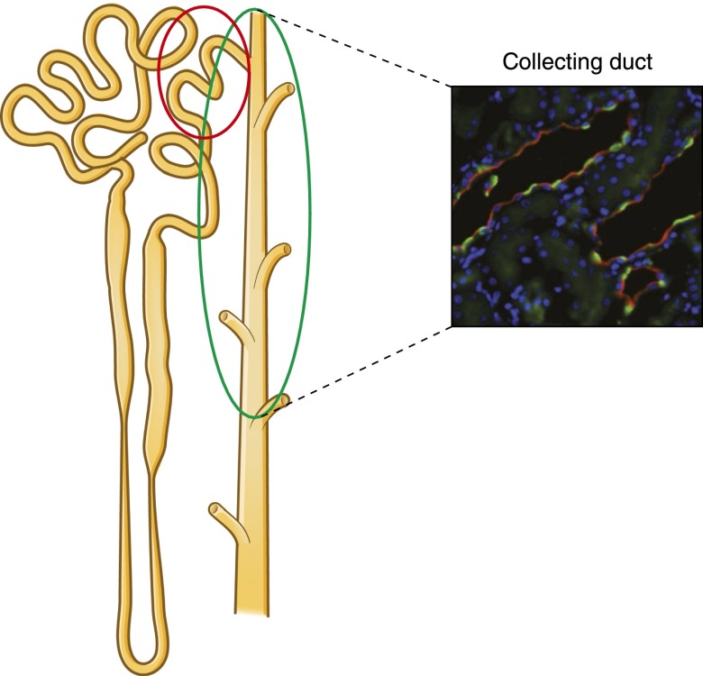
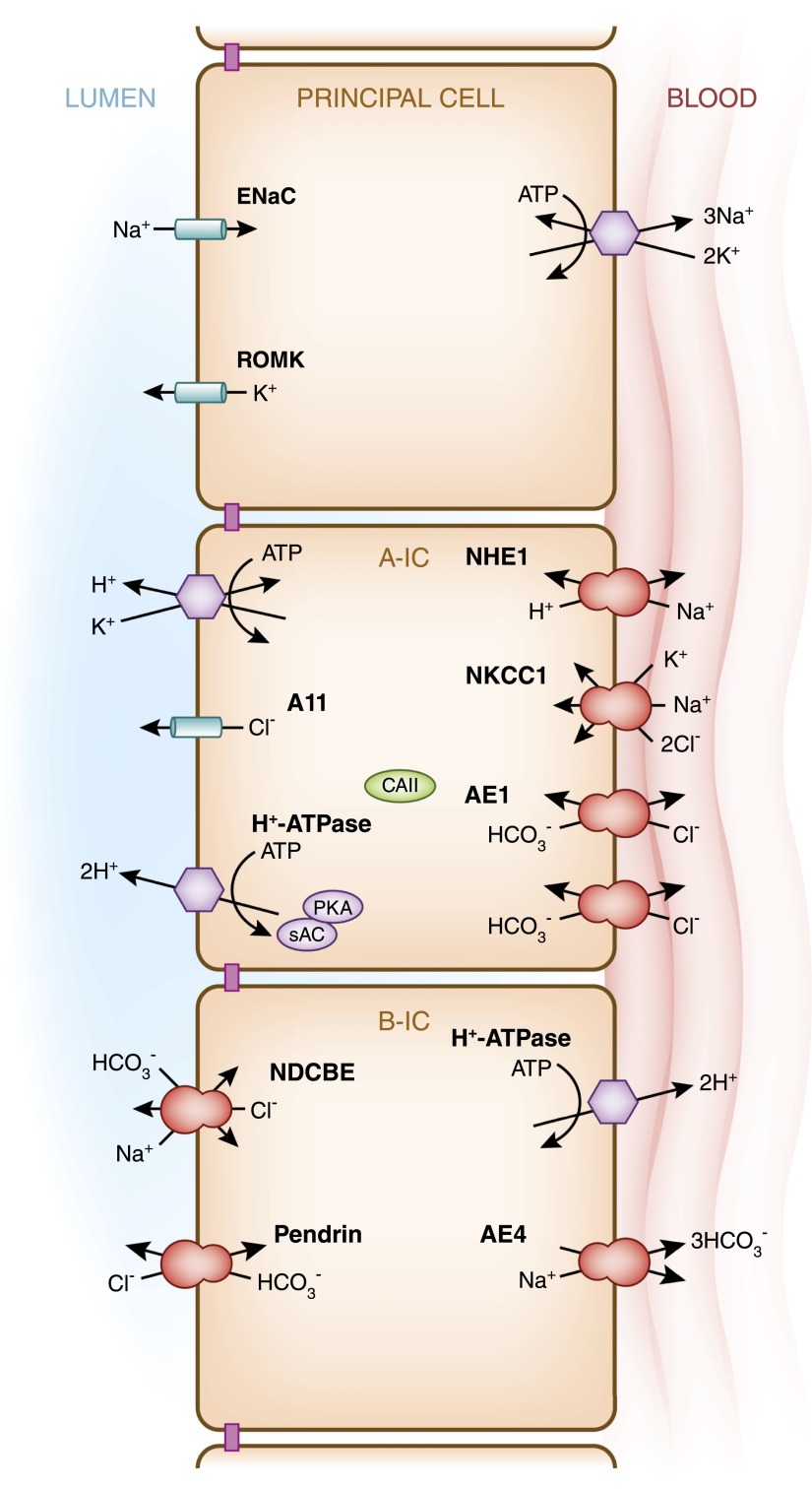
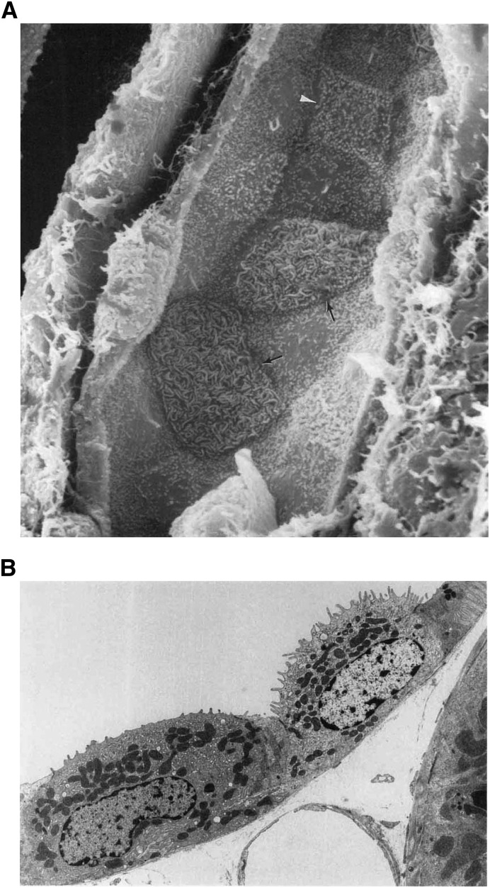
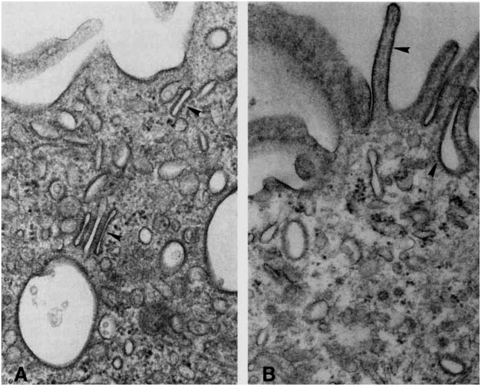
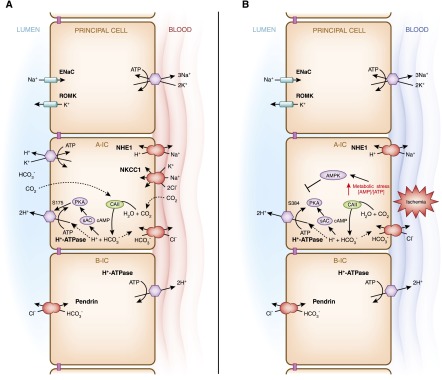
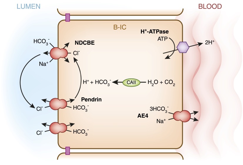
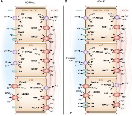
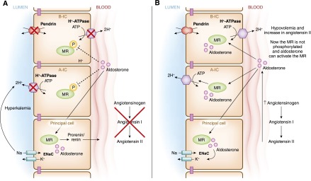
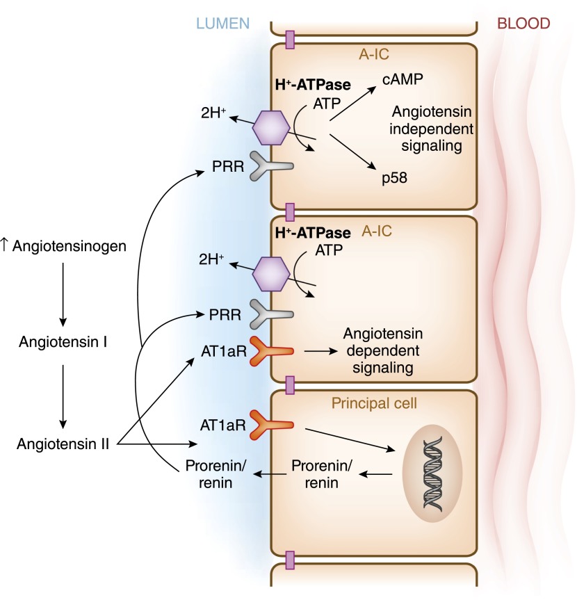
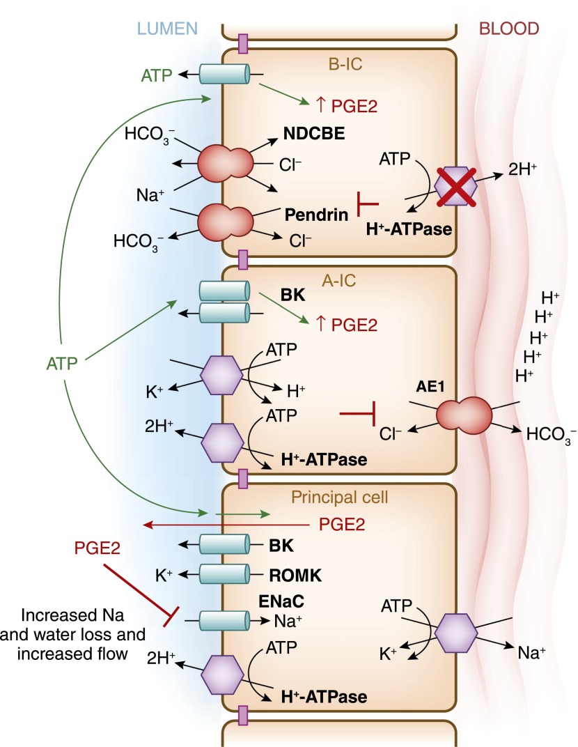
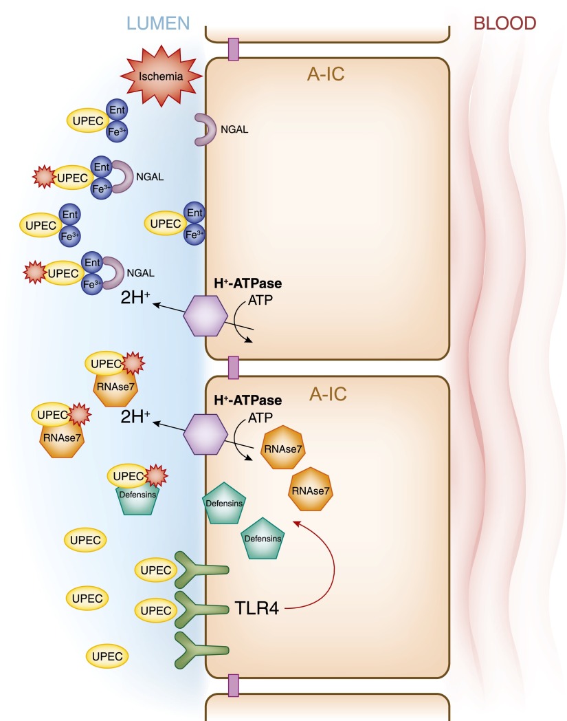
Intercalated cells are kidney tubule epithelial cells with important roles in the regulation of acid-base homeostasis. However, in recent years the understanding of the function of the intercalated cell has become greatly enhanced and has shaped a new model for how the distal segments of the kidney tubule integrate salt and water reabsorption, potassium homeostasis, and acid-base status. These cells appear in the late distal convoluted tubule or in the connecting segment, depending on the species. They are most abundant in the collecting duct, where they can be detected all the way from the cortex to the initial part of the inner medulla. Intercalated cells are interspersed among the more numerous segment-specific principal cells. There are three types of intercalated cells, each having distinct structures and expressing different ensembles of transport proteins that translate into very different functions in the processing of the urine. This review includes recent findings on how intercalated cells regulate their intracellular milieu and contribute to acid-base regulation and sodium, chloride, and potassium homeostasis, thus highlighting their potential role as targets for the treatment of hypertension. Their novel regulation by paracrine signals in the collecting duct is also discussed. Finally, this article addresses their role as part of the innate immune system of the kidney tubule.

Keywords: aldosterone; blood pressure; cell and transport; distal tubule; physiology; renal tubular acidosis.


Figures

Figure 1.
Figure 1.
Intercalated cells in rat collecting duct. The cartoon and confocal micrograph illustrate the intercalated cell distribution along the kidney tubule and within the epithelium. Intercalated cells were detected in the cortical and outer medullary collecting duct (green oval) by immunoflorescence labeling using an antibody against one of the H+-ATPase subunits (green). Intercalated cells are also located in the connecting segment (red circle). The apical or luminal labeling of the H+-ATPase indicates that these cells are mostly type A intercalated cells (A-IC). Principal cells were labeled with an antibody against the water channel aquaporin-2 (red). Modified from reference , with permission.
Figure 2.
Figure 2.
Transepithelial transport processes and regulatory mechanisms in type A intercalated cells (A-IC) and type B intercalated cells (B-IC). This cartoon illustrates the major transport proteins expressed in the three main epithelial cell types present in the collecting duct: the principal cell, which expresses the epithelial sodium channel; the acid-secreting type A-IC; and type B-IC, which secretes bicarbonate while reabsorbing NaCl. In the cortical and outer medullary collecting duct, type A-ICs express H+-ATPase and the H+/K+-ATPase at the apical/luminal membrane, while they express the Cl/HCO3 exchanger AE1 at their basolateral membrane. The bicarbonate sensor soluble adenylyl cyclase (sAC) and protein kinase A (PKA) play important roles in the regulation of the H+-ATPase (see Figure 5A). Slc26a11 (A11), an electrogenic Cl transporter, as well as a Cl/HCO3 anion exchanger, are also expressed at the apical membrane of the type A-IC. On the other hand, the type B-ICs display an electroneutral NaCl transport/reabsorption pathway at their apical membrane that involves pendrin, a Cl/HCO3 exchanger, and the Na+-driven Cl/HCO3 exchanger (NDCBE). The proposed basolateral Na+ extrusion pathway would involve the cotransporter Slc4a9 (AE4). The mechanism of Cl exit remains to be elucidated. In type B-ICs, reabsorption of NaCl from the lumen is energized by the basolateral H+-ATPase rather than by Na+/K+-ATPase.
Figure 3.
Figure 3.
Morphology of rat cortical collecting duct intercalated cells. (A) The scanning electron micrograph shows the luminal surface of the rat collecting duct. In this image principal cells can be easily identified by their small microprojections into the lumen and by the presence of a single cilium. Two configurations of intercalated cells are present in this tubule. The type A-ICs (arrows) have a large luminal surface covered mostly with microplicae, while a type B-IC (arrowhead) has a more angular cellular outline and smaller apical microvilli (original magnification, ×5500). Reproduced with permission from reference 10. (B) This transmission electron micrograph of rat cortical collecting duct illustrates further the two configurations of intercalated cells. The type A-IC (right) shows a well developed apical tubulovesicular membrane compartment, with prominent microprojections that are part of the microplicae on the luminal membrane. In this image, the type B-IC (left) presents a denser cytoplasm with more abundant mitochondria and many vesicles throughout the cytoplasm. The luminal membrane of the type B cell has a quite smooth luminal membrane (original magnification, ×5000). Reproduced with permission from reference .
Figure 4.
Figure 4.
Change in intercalated cell morphology in response to chronic acid-base status changes. This transmission electron micrograph shows the apical membrane of type A cells from the collecting duct from a normal rat (A) and from a rat with acute respiratory acidosis (B). In the control animal prominent studs (arrowheads) are observed on tubulovesicular structures in the control rat (A), while they are more abundant on the apical membrane in the experimental animal (B). In another study, Brown and colleagues showed that these studs are H+-ATPase, both at the membrane and in the tubulovesicular subapical structures (57). The increase in the number of H+-ATPases (studs) at the apical membrane in the animal with acidosis (B) is coupled to an increase in membrane microprojections and a decrease in the number of tubulovesicular structures in the acidotic rat (B). (Original magnification: A, ×48,000; B, ×42,400.) Reproduced with permission from reference .
Figure 5.
Figure 5.
Model of H+-ATPase coregulation at the apical membrane of type A intercalated cells by two kinases, downstream of acid-base status and of cellular metabolic stress. (A) Acute increases in the level of intracellular bicarbonate activates the bicarbonate-sensor sAC, which generates cAMP and then activates PKA. Studies using pharmacological activators have shown that exchange protein directly activated by cAMP (Epac) is not likely to play a role in H+-ATPase regulation (183). Carbonic anhydrase II (CAII) is involved in the generation of intracellular bicarbonate. Downstream of PKA, the A subunit of H+-ATPase is then phosphorylated at Ser-175 (S175) (72). This phosphorylation event is involved in activating H+-ATPase at the apical membrane. (B) This acute stimulatory effect of the sAC/cAMP/PKA signaling cascade on apical H+-ATPase activity is counterbalanced by the inhibitory effect of the metabolic sensor AMP activated protein kinase (AMPK), downstream of ischemia or metabolic stress, for example. Acute metabolic stress leads to an elevation of the levels of AMP compared with ATP, and the increase in cellular [AMP]/[ATP] directly activates AMPK. AMPK mediates the downregulation of H+-ATPase activity by phosphorylating Ser-384 (S384) in the proton pump’s A subunit (73).
Figure 6.
Figure 6.
Model of major transport processes and regulatory mechanisms in type B intercalated cells. This cartoon is modified from the model proposed by Chambrey and colleagues (. These cells participate in electroneutral NaCl absorption, which is energized by H+-ATPase. Two cycles of transport via pendrin, when coordinated with one cycle of NDCBE results in net uptake of one Na+ and one Cl and the extrusion of two bicarbonate (HCO3) ions. The Cl ions recycle across the apical membrane. A basolateral channel (not shown) mediates Cl exit. The basolateral anion exchanger 4 (AE4 or Slc4a9), together with the H+-ATPase also facilitates Na+ and bicarbonate exit.
Figure 7.
Figure 7.
Intercalated cells are involved in K+ secretion in the collecting duct. This cartoon illustrates the location of the voltage-dendent renal outer medullary small-conductance K+ (ROMK) and the flow-dependent big potassium (BK) channels in the intercalated cells (type A-IC and type B-IC) and principal cells. (A) Under normal dietary conditions, ROMK predominantly secretes K+. However, under the influence of a K+-rich diet, because of the increased activity of the basolateral Na+/K+-ATPase, the transport via the epithelial sodium channel (ENaC)– transport increases, generating a driving force for ROMK to secrete more K+ from the principal cells. (B) When high K+ secretion is accompanied by increased flow, it stimulates BK channel synthesis and function in the intercalated cells (type A-IC and type B-IC).
Figure 8.
Figure 8.
Model of regulatory pathways for the mineralocorticoid receptor (MR) in intercalated cells: hyperkalemia and volume depletion. This figure depicts pathways that involve the differential phosphorylation state of the MR in principal versus intercalated cells (type A-IC; type B-IC), that are likely the key to the distinct responses of these cells in two different scenarios. (A) The first scenario involves conditions when only aldosterone is present (as in hyperkalemia). In this case, hyperkalemia leads to aldosterone secretion while no angiotensin II is present. Here, MR is phosphorylated in intercalated but not in principal cells. These conditions lead to aldosterone-mediated Na+ reabsorption via the epithelial sodium channel in principal cells, which drives K+ secretion also in principal cells. (B) In contrast, when both angiotensin II and aldosterone are present (as in intravascular volume depletion), the MR is dephosphorylated downstream of angiotensin II, and the activity of this receptor is thus restored in intercalated cells. In addition, as a result of aldosterone signaling both pendrin and H+-ATPase are upregulated, and in turn there is a decrease drive for K+ secretion. Modified from reference .
Figure 9.
Figure 9.
Intercalated cells are targets of paracrine angiotensin II signaling. Angiotensin II stimulates secretion of prorenin and renin from principal cells. Prorenin binds to the prorenin receptor at the apical membrane of type A-IC, where in turn it stimulates the synthesis of angiotensin II from angiotensin I via the action of angiotensinogen, which originates in proximal segments of the nephron. Angiotensin II then binds to the angiotensin receptor 1a (AT1aR) in the type A-IC. Moreover, prorenin binds to its receptor in type A-IC to stimulate signaling via p58 or cAMP.
Figure 10.
Figure 10.
Intercalated cells are necessary to maintain body fluid and electrolyte balance. This model summarizes the recent findings by Gueutin and colleagues (35), which showed how H+-ATPase dysfunction in type B-IC leads to the urinary water and sodium losses in patients with distal renal tubule acidosis. This group studied mice with significantly decreased H+-ATPase activity in the collecting duct intercalated cells due to knockout of the B1 of the H+-ATPase subunit (ATP6V1B1–/–mice). The specific knockout of the B1 subunit does not disturb H+-ATPase expression in the proximal tubule. These animals presented with a significant urinary loss of NaCl, revealing impaired function of epithelial sodium channel (ENaC) in principal cells, as well as decreased pendrin and NDCBE function in type B-IC in the cortical collecting duct. These animals had an upregulation of ENaC in the medulla. High levels of prostaglandin E2 (PGE2) and ATP were detected in the urine of these animals. When PGE2 was normalized using pharmacologic agents these animals also normalized their ENaC levels in the cortex, and they had improved polyuria and hypokalemia. When the H+-ATPase was inactivated in type B-IC, it resulted in ATP release with the subsequent increases in PGE2 release from these cell as well.
Figure 11.
Figure 11.
The intercalated cells as key effectors of the innate immune system. This model presents various signaling cascades triggered in intercalated cells by pathologic conditions that can result in the release of defensins. Collecting duct epithelial cells respond to gram-negative bacteria (uropathogenic Escherichia coli [UPEC]) by activating cascades drownstream from the toll-like receptor 4 (TLR4) and some TLR4-independent pathways (lower). Ischemic injury also induces release of the defensin neutrophil gelatinase-associated lipocalin from intercalated cells (NGAL) (upper), and type A-IC also secrete the defensin RNAase7, which is a key molecule to prevent urinary tract infection. Funtional apical H+-ATPase is also relevant for the type A intercalated cell anti-microbial function.

References

    1. Crayen ML, Thoenes W: Architecture and cell structures in the distal nephron of the rat kidney. Cytobiologie 17: 197–211, 1978 - PubMed
    1. Davies JA, Davey MG: Collecting duct morphogenesis. Pediatr Nephrol 13: 535–541, 1999 - PubMed
    1. Meneton P, Loffing J, Warnock DG: Sodium and potassium handling by the aldosterone-sensitive distal nephron: The pivotal role of the distal and connecting tubule. Am J Physiol Renal Physiol 287: F593–F601, 2004 - PubMed
    1. Eladari D, Chambrey R, Peti-Peterdi J: A new look at electrolyte transport in the distal tubule. Annu Rev Physiol 74: 325–349, 2012 - PMC - PubMed
    1. Madsen KM, Tisher CC: Structural-functional relationship along the distal nephron. Am J Physiol 250: F1–F15, 1986 - PubMed

Publication types