. 2015 Jan 28;35(4):1530-8.
doi: 10.1523/JNEUROSCI.2278-14.2015.

Lymphocytes from chronically stressed mice confer antidepressant-like effects to naive mice

Affiliations

Lymphocytes from chronically stressed mice confer antidepressant-like effects to naive mice

Rebecca A Brachman et al. J Neurosci. .

Abstract

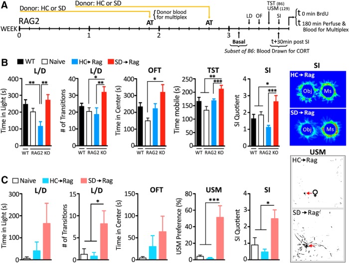
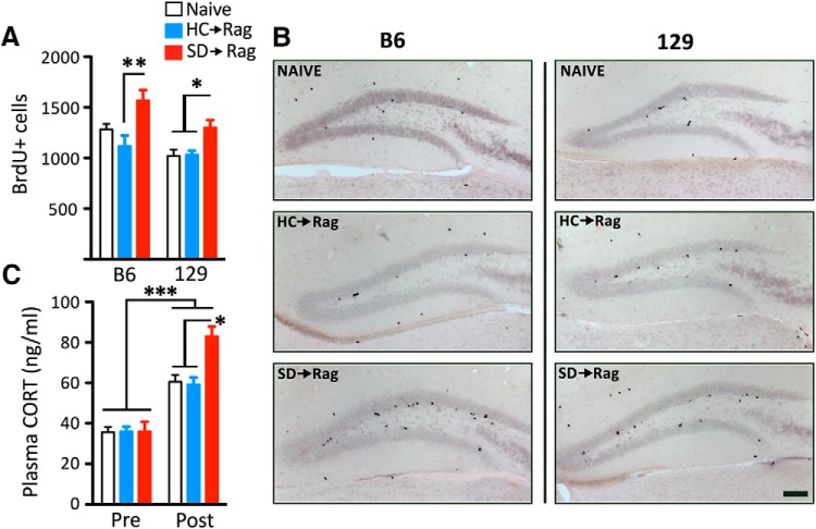
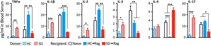
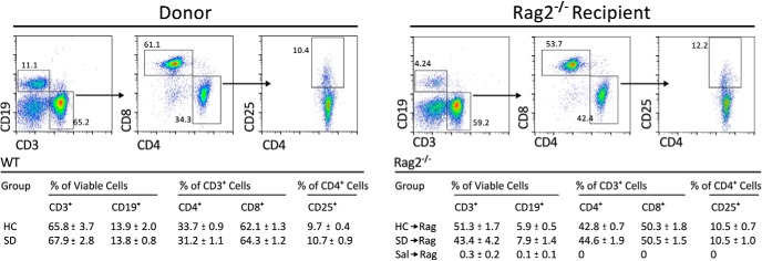
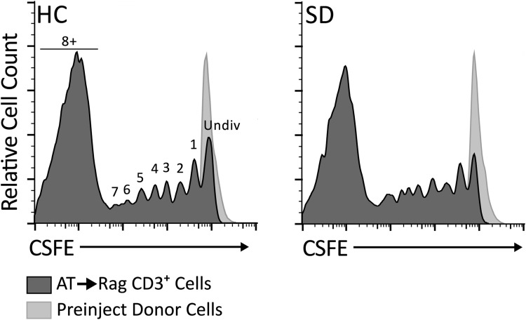
We examined whether cells of the adaptive immune system retain the memory of psychosocial stress and thereby alter mood states and CNS function in the host. Lymphocytes from mice undergoing chronic social defeat stress or from unstressed control mice were isolated and adoptively transferred into naive lymphopenic Rag2(-/-) mice. Changes in affective behavior, hippocampal cell proliferation, microglial activation states, and blood cytokine levels were examined in reconstituted stress-naive mice. The mice receiving lymphocytes from defeated donors showed less anxiety, more social behavior, and increased hippocampal cell proliferation compared with those receiving no cells or cells from unstressed donors. Mice receiving stressed immune cells had reduced pro-inflammatory cytokine levels in the blood relative to the other groups, an effect opposite to the elevated donor pro-inflammatory cytokine profile. Furthermore, mice receiving stressed immune cells had microglia skewed toward an anti-inflammatory, neuroprotective M2-like phenotype, an effect opposite the stressed donors' M1-like pro-inflammatory profile. However, stress had no effect on lymphocyte surface marker profiles in both donor and recipient mice. The data suggest that chronic stress-induced changes in the adaptive immune system, contrary to conferring anxiety and depressive behavior, protect against the deleterious effects of stress. Improvement in affective behavior is potentially mediated by reduced peripheral pro-inflammatory cytokine load, protective microglial activity, and increased hippocampal cell proliferation. The data identify the peripheral adaptive immune system as putatively involved in the mechanisms underlying stress resilience and a potential basis for developing novel rapid-acting antidepressant therapies.

Keywords: adaptive immune system; cytokines; depression; hippocampal neurogenesis; lymphocytes; stress.


Figures

Figure 1.
Figure 1.
Transfer of lymphocytes from stressed mice confers behavioral resilience in two strains of Rag2−/− mice. The experimental time line is shown in A. Following adoptive transfer (AT) of cells from SD or HC donors, SD→Rag mice on a C57BL/6 background (n = 12–16 per group; B) showed increased time in light and elevated transitions in the L/D box, increased center time in the OFT, increased mobility in the TST, and enhanced preference for SI. Behavioral profiles between C57BL/6 WT and naive Rag2−/− mice on the same background were comparable. SD→Rag mice on 129 background (n = 6–8 per group; C) showed comparable anxiolytic changes in the L/D test and significantly enhanced expressions of socially affiliative behaviors measured in the USM and SI tests. Post hoc significances marked. *p < 0.5; **p < 0.01; ***p < 0.001. Heat maps show digitized behavior in the SI task, and binarized maps show marking behavior to female urine in the USM test.
Figure 2.
Figure 2.
Transfer of lymphocytes from stressed mice increases hippocampal DG cell proliferation in the C57BL/6 (A) and 129 (B) strains of Rag2−/− mice and increases SI-induced corticosterone (CORT) levels in C57BL/6 mice (n = 10–16 per group; C). The increases in SD→Rag mice were relative to naive and HC→Rag mice with significances indicated. Representative photomicrographs of the BrdU-labeled cells in the DG of the two strains are shown. Scale bar, 100 μm.
Figure 3.
Figure 3.
Levels of several plasma cytokines are increased by SD stress relative to HC control mice, but the relationship is reversed in Rag2−/− mice receiving cells from those animals. In HC→Rag mice elevations relative to naive control were seen for some cytokines. In the SD→Rag mice, reductions in levels were seen for pro-inflammatory cytokines and increases in levels of the anti-inflammatory cytokine IL-4 were seen relative to the other groups, with significances shown (n = 4–6 per group).
Figure 4.
Figure 4.
The relative abundance of lymphocyte subpopulations was not affected by SD stress compared with HC control conditions (n = 6 per group). Representative bivariate dot plots show lymphocytes from donor mice and host Rag2−/− mice (examples are from lymph nodes taken from mice in defeat conditions) stained and gated for B cells, T cells, and T cell subsets. The table shows that ratios of CD19+, CD3+, CD4+, and CD8+ cells as well as the proportion of CD25+ regulatory CD4+ T cells were not affected.
Figure 5.
Figure 5.
Cell-proliferation rates of adoptively transferred (AT) cells in the Rag2−/− mice were not affected by SD stress in the donor mice. The CFSE intensity histograms show that most cells underwent >8 divisions in the period after the two transfers (8 and 14 d prior). Two representative histograms are shown for lymph node cells from HC→Rag and SD→Rag mice. Successive cell divisions to the left of the undivided peak (obtained from CFSE-labeled donor cells) are labeled 1–7 and 8+. Though rates of proliferation trended higher in SD→Rag samples, t test of cells in the undivided (Undiv) peak between HC and SD was p = 0.06 (n = 3–4), and no other differences were significant.

References

    1. Battista D, Ferrari CC, Gage FH, Pitossi FJ. Neurogenic niche modulation by activated microglia: transforming growth factor β increases neurogenesis in the adult dentate gyrus. Eur J Neurosci. 2006;23:83–93. doi: 10.1111/j.1460-9568.2005.04539.x. - DOI - PubMed
    1. Beurel E, Harrington LE, Jope RS. Inflammatory T helper 17 cells promote depression-like behavior in mice. Biol Psychiatry. 2013;73:622–630. doi: 10.1016/j.biopsych.2012.09.021. - DOI - PMC - PubMed
    1. Brynskikh A, Warren T, Zhu J, Kipnis J. Adaptive immunity affects learning behavior in mice. Brain Behav Immun. 2008;22:861–869. doi: 10.1016/j.bbi.2007.12.008. - DOI - PubMed
    1. Cherry JD, Olschowka JA, O'Banion MK. Neuroinflammation and M2 microglia: the good, the bad, and the inflamed. J Neuroinflammation. 2014;11:98. doi: 10.1186/1742-2094-11-98. - DOI - PMC - PubMed
    1. Cho BK, Rao VP, Ge Q, Eisen HN, Chen J. Homeostasis-stimulated proliferation drives naive T cells to differentiate directly into memory T cells. J Exp Med. 2000;192:549–556. doi: 10.1084/jem.192.4.549. - DOI - PMC - PubMed

Publication types

MeSH terms