. 2015 Jan 28;35(4):1781-91.
doi: 10.1523/JNEUROSCI.2998-14.2015.

Morphometric and histologic substrates of cingulate integrity in elders with exceptional memory capacity

Affiliations

Morphometric and histologic substrates of cingulate integrity in elders with exceptional memory capacity

Tamar Gefen et al. J Neurosci. .

Abstract

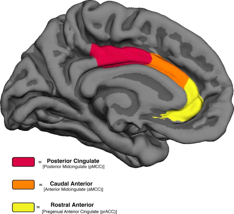
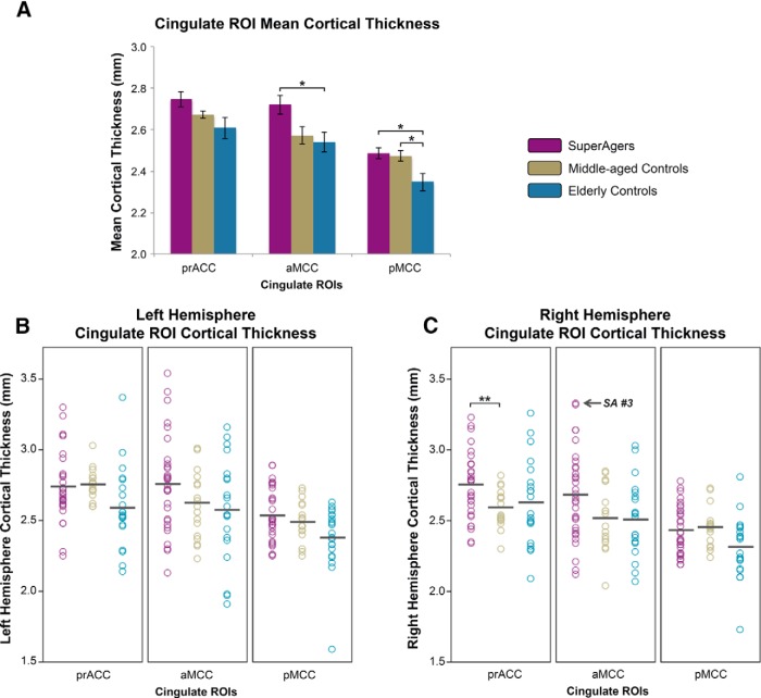
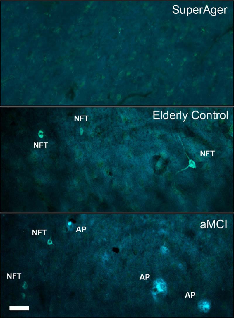
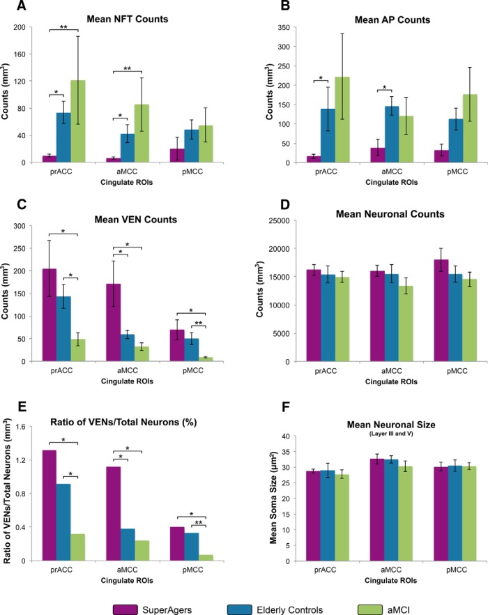
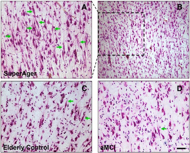
This human study is based on an established cohort of "SuperAgers," 80+-year-old individuals with episodic memory function at a level equal to, or better than, individuals 20-30 years younger. A preliminary investigation using structural brain imaging revealed a region of anterior cingulate cortex that was thicker in SuperAgers compared with healthy 50- to 65-year-olds. Here, we investigated the in vivo structural features of cingulate cortex in a larger sample of SuperAgers and conducted a histologic analysis of this region in postmortem specimens. A region-of-interest MRI structural analysis found cingulate cortex to be thinner in cognitively average 80+ year olds (n = 21) than in the healthy middle-aged group (n = 18). A region of the anterior cingulate cortex in the right hemisphere displayed greater thickness in SuperAgers (n = 31) compared with cognitively average 80+ year olds and also to the much younger healthy 50-60 year olds (p < 0.01). Postmortem investigations were conducted in the cingulate cortex in five SuperAgers, five cognitively average elderly individuals, and five individuals with amnestic mild cognitive impairment. Compared with other subject groups, SuperAgers showed a lower frequency of Alzheimer-type neurofibrillary tangles (p < 0.05). There were no differences in total neuronal size or count between subject groups. Interestingly, relative to total neuronal packing density, there was a higher density of von Economo neurons (p < 0.05), particularly in anterior cingulate regions of SuperAgers. These findings suggest that reduced vulnerability to the age-related emergence of Alzheimer pathology and higher von Economo neuron density in anterior cingulate cortex may represent biological correlates of high memory capacity in advanced old age.

Keywords: Alzheimer's pathology; aging; cingulate cortex; cognition; histology; structural MRI.


Figures

Figure 1.
Figure 1.
Three cingulate ROIs. Medial ROIs of the cingulate cortex in the Desikan-Killiany (Desikan et al., 2006) cortical labeling protocol are color-coded with their corresponding parcellations characterized by Vogt (2009).
Figure 2.
Figure 2.
A–C, Cingulate ROI cortical thickness from superagers, middle-aged controls, and elderly controls. A, Bar graphs demonstrate the distribution of mean (average of left and right hemisphere) cortical thickness values in SuperAgers, middle-aged controls, and elderly controls and SE bars are shown. B, C, Scatterplots show individual cortical thickness measures in left hemisphere (LH) and right hemisphere (RH) in all three ROIs across subject groups. Black bars indicate means of each scatterplot column. SuperAger 3 is indicated to emphasize high caudal anterior thickness. *p < 0.05; **p < 0.01.
Figure 3.
Figure 3.
Thioflavin-S staining of NFTs and APs at 20× magnification. The photomicrographs above show no (or extremely sparse) Alzheimer neuropathology (APs and NFTs) in the anterior aspects of cingulate cortex (aMCC) in a 90-year-old SuperAger (SuperAger 3) compared with an age-matched elderly control (middle), and an individual diagnosed clinically with aMCI (bottom). Scale bar in bottom left corner indicates 50 μm at 20×.
Figure 4.
Figure 4.
A–F, Heights of the bars represent mean estimated counts of NFTs, APs, VENs, total neurons, and size of neurons, respectively, per cubic millimeter in subject groups (n = 5 per group). A, B, Mean numerical estimates of NFT and AP density were lowest in SuperAgers, followed by the elderly control and aMCI groups across all cingulate regions, with the exception of AP density in aMCC. Significantly lower NFT density in anterior cingulate regions (prACC and aMCC) in SuperAgers compared with the other groups were found, whereas AP density was more variable, with differences reaching statistical significance between SuperAgers and elderly controls in anterior cingulate regions. C, The inverse pattern was demonstrated in VEN counts across regions, where SuperAgers showed highest counts, especially in anterior cingulate regions (aMCC), followed by elderly controls and aMCI individuals. D, There were no differences in total neuronal counts across the subject groups. E, When total neuronal count was accounted for, patterns of VEN distributions (illustrated as the ratio of VEN counts to total neuronal counts) remained similar to the distribution illustrated in VEN counts alone (C); this is due to generally equal counts of total neurons within regions and across groups. F, No differences in total mean neuronal size (area is in mean squared micrometers) across groups were found. *p < 0.05; **p < 0.01.
Figure 5.
Figure 5.
A–D, Nissl stain at 20× magnification in aMCC shows higher density of VENS (green arrows) in a SuperAger (A, SuperAger 3) compared with an elderly control (C) and an individual with aMCI (D). Photomicrograph B displays very densely packed VENs in SuperAger 3, clearly visible in abundance at 10× magnification; selected dashed region highlights the same region that is displayed in higher power in 5A. Scale bar in bottom right corner indicates 50 μm in photomicrographs A, C, and D at 20×, and 100 μm in photomicrograph B at 10×.

References

    1. Albert MS, DeKosky ST, Dickson D, Dubois B, Feldman HH, Fox NC, Gamst A, Holtzman DM, Jagust WJ, Petersen RC, Snyder PJ, Carrillo MC, Thies B, Phelps CH. The diagnosis of mild cognitive impairment due to Alzheimer's disease: recommendations from the National Institute on Aging-Alzheimer's Association workgroups on diagnostic guidelines for Alzheimer's disease. Alzheimers Dement. 2011;7:270–279. doi: 10.1016/j.jalz.2011.03.008. - DOI - PMC - PubMed
    1. Allman JM, Hakeem A, Erwin JM, Nimchinsky E, Hof P. The anterior cingulate cortex. Ann N Y Acad Sci. 2001;935:107–117. doi: 10.1111/j.1749-6632.2001.tb03476.x. - DOI - PubMed
    1. Allman JM, Tetreault NA, Hakeem AY, Manaye KF, Semendeferi K, Erwin JM, Park S, Goubert V, Hof PR. The von Economo neurons in frontoinsular and anterior cingulate cortex in great apes and humans. Brain Structure and Function. 2010;214:495–517. doi: 10.1007/s00429-010-0254-0. - DOI - PubMed
    1. Arriagada PV, Growdon JH, Hedley-Whyte ET, Hyman BT. Neurofibrillary tangles but not senile plaques parallel duration and severity of Alzheimer's disease. Neurology. 1992;42:631–639. doi: 10.1212/WNL.42.3.631. - DOI - PubMed
    1. Balasubramanian AB, Kawas CH, Peltz CB, Brookmeyer R, Corrada MM. Alzheimer disease pathology and longitudinal cognitive performance in the oldest-old with no dementia. Neurology. 2012;79:915–921. doi: 10.1212/WNL.0b013e318266fc77. - DOI - PMC - PubMed

Publication types

MeSH terms

Substances