

Pediatric synovial sarcoma in the retropharyngeal space: a rare and unusual presentation
- PMID: 25632364
- PMCID: PMC4302351
- DOI: 10.1155/2015/587386
Pediatric synovial sarcoma in the retropharyngeal space: a rare and unusual presentation
Abstract
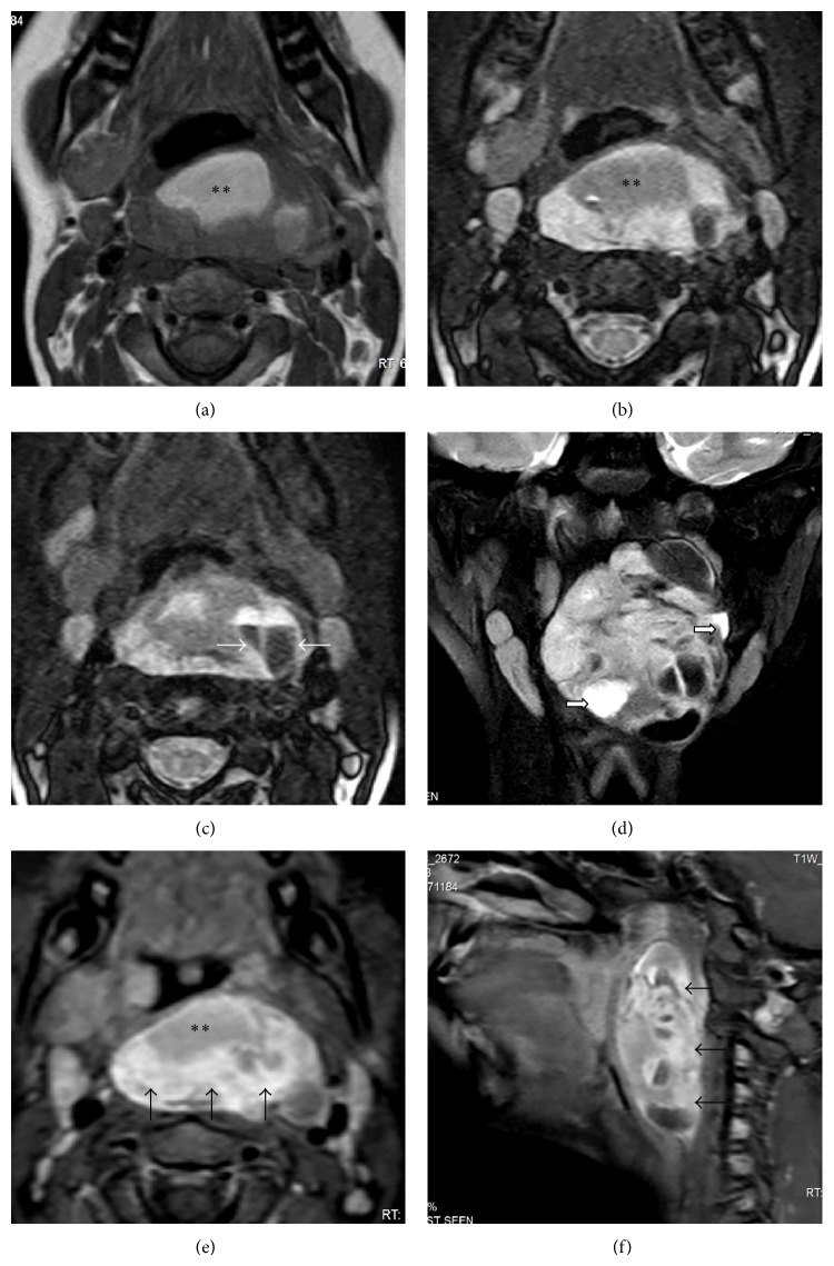
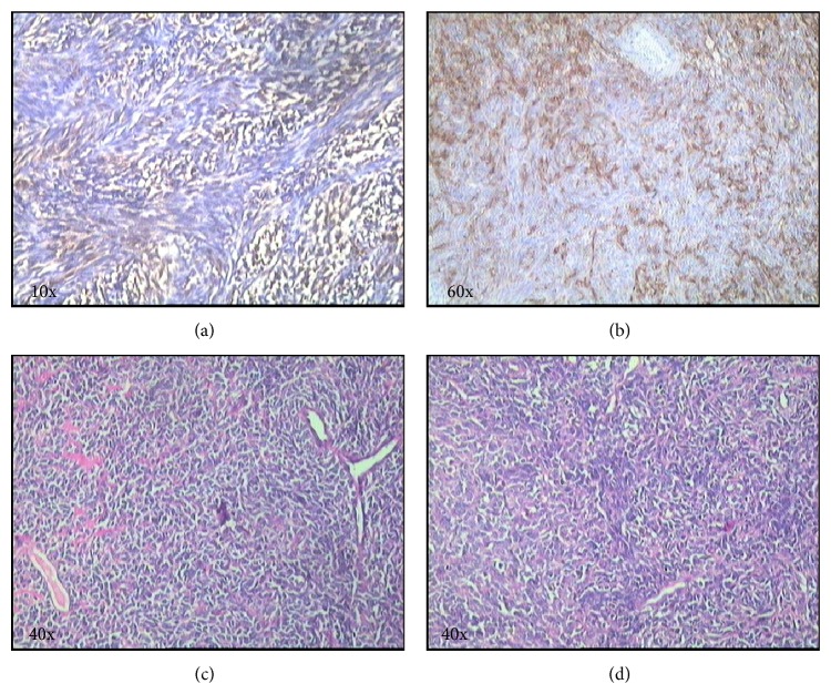
Synovial sarcomas in the head and neck are extremely rare tumors, especially in the pediatric population. 3-5% of synovial sarcomas occur in the head and neck region displaying varied imaging and histopathological features resulting in frequent misdiagnosis. These tumors have a poor prognosis; hence early diagnosis and accurate classification based on imaging, histopathology, and immunohistochemistry are critical for prompt treatment. To the best of our knowledge, imaging findings of pediatric retropharyngeal lipomatous synovial sarcoma have not been reported to date in English medical literature. We report, for the first time, a rare case of retropharyngeal lipomatous synovial sarcoma in a ten-year-old child and discuss the case-specific imaging findings in our patient using magnetic resonance imaging and computed tomography.
Figures
Similar articles
-
Synovial sarcoma of the hypopharynx in a pediatric patient: Case report.Int J Surg Case Rep. 2016;28:1-3. doi: 10.1016/j.ijscr.2016.08.043. Epub 2016 Sep 3. Int J Surg Case Rep. 2016. PMID: 27649458 Free PMC article.
-
[Synovial sarcoma of the head and neck: two cases report].Rev Laryngol Otol Rhinol (Bord). 2005;126(1):53-6. Rev Laryngol Otol Rhinol (Bord). 2005. PMID: 16080650 French.
-
Primary pericardial synovial sarcoma in an adolescent patient: magnetic resonance and diffusion-weighted imaging features.J Pediatr Hematol Oncol. 2015 May;37(4):e230-3. doi: 10.1097/MPH.0000000000000305. J Pediatr Hematol Oncol. 2015. PMID: 25647483
-
Lipoblastomatosis of the retropharyngeal space: pathogenesis, presentation, and management, with a focus on head-neck lipoblastoma(toses).B-ENT. 2016;12(1):33-9. B-ENT. 2016. PMID: 27097392 Review.
-
Retropharyngeal synovial sarcoma in an infant: report of a case and of its response to chemotherapy; review of the literature.Pediatr Hematol Oncol. 1991 Jan-Mar;8(1):45-52. doi: 10.3109/08880019109033426. Pediatr Hematol Oncol. 1991. PMID: 2029466 Review.
References
-
- Pugliese G. N., Saetti R., Furlanetto A. Synovial sarcoma of the head and neck: a case report of parapharyngeal region and review of the literature. Acta Otorhinolaryngologica Italica. 1992;12(4):389–397. (Ita). - PubMed
-
- Yoshihara T., Kawano K., Mita N. Retropharyngeal lipoma causing severe dysphagia and dyspnea. Journal of Otolaryngology. 1998;27(6):363–366. - PubMed
-
- Yueh B., Bassewitz H. L., Eisele D. W. Retropharyngeal liposarcoma. The American Journal of Otolaryngology. 1995;16(5):331–340. - PubMed
LinkOut - more resources
Full Text Sources
Other Literature Sources
