. 2015 Jun;57(6):506-12.
doi: 10.1007/s12033-015-9844-7.

In-Cell RNA Hydrolysis Assay: A Method for the Determination of the RNase Activity of Potential RNases

Affiliations

In-Cell RNA Hydrolysis Assay: A Method for the Determination of the RNase Activity of Potential RNases

Youngsil Seo et al. Mol Biotechnol. 2015 Jun.

Abstract

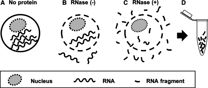
Conventional procedures to assay RNA degradation by a protein with ribonuclease (RNase) activity require a step to isolate intact RNA molecules, which are used as a substrate. Here, we established a novel "In-cell RNA hydrolysis assay" in which RNAs within cells are used as a substrate for the RNA-hydrolyzing protein, thereby avoiding the need to prepare intact RNA molecules. In this method, the degree of RNA degradation is indicated by the fluorescence intensity of RNA molecules released from fixed and permeabilized cells following treatment with the potential RNase. A catalytic 3D8 antibody capable of degrading RNAs and pancreatic RNase A were used as model RNases. Our data demonstrate that the novel In-cell RNA hydrolysis assay is a reliable and sensitive method to analyze the activities of potential RNA-hydrolyzing proteins such as catalytic antibodies.


Figures

Fig. 1
Fig. 1
Schematic representation of the concept of the In-cell RNA hydrolysis assay. a Cells containing intact RNAs. b Cells whose membranes have been permeabilized, allowing the release of small RNA fragments even in the absence of RNase treatment. c Cells release small RNA fragments via their membrane pores following treatment with an RNase. d Collection of degraded RNA fragments released from cells, which are labeled with RiboGreen and subjected to fluorescence intensity measurement
Fig. 2
Fig. 2
The detection of RNAs released from fixed and permeabilized cells. a Experimental procedures for the data presented in (b) and (d). b A549 cells grown in a 48-well plate at a density of 5 × 104 cells/well were fixed, permeabilized, and incubated with 100 μl of 10 μM RNase A or 10 μM 3D8 antibody for 2 h at 37 °C. An aliquot of the conditioned medium was added to RiboGreen, and the fluorescence intensity was analyzed. c Pure 16S and 23S rRNA from E. coli was incubated with RiboGreen in the presence or absence of RNase A prior to fluorescence intensity analysis. d A549 cells grown in a 6-well plate at a density of 5 × 105 cells/well were fixed, permeabilized, and incubated with 10 μM RNase A or 3D8 antibody for 2 h at 37 °C. Proteins were removed from the conditioned medium by precipitation, and absorbance at 260 nm was measured. e Degradation of plasmid DNA (1 μg/ml) by DNase I (2 U) was tested using RiboGreen prepared in DNase I reaction buffer for 2 h at 37 °C. f Fixed and permeabilized cells in a 48-well plate were treated with RNase A or 3D8 antibody (10 μM) in the presence or absence of DNase I (2 U) for 2 h at 37 °C. The conditioned medium was mixed with RiboGreen prior to fluorescence intensity analysis. RFU relative fluorescence unit. Data represent the mean ± standard error of four independent experiments
Fig. 3
Fig. 3
Quantitative analysis of RNA degradation in fixed and permeabilized cells treated with RNase A or 3D8 antibody. a Standard curve to determine the concentration of RNA. Pure rRNA at known concentrations was mixed with RiboGreen prior to fluorescence intensity analysis. b Dose-dependent RNA degradation. RNA degradation was quantitatively analyzed in fixed and permeabilized cells (5 × 104 cells) treated with 1.25–10 μM RNase A or 3D8 antibody. c The sensitivity of this method. RNA degradation was quantitatively analyzed in fixed and permeabilized cells treated with various dilutions of RNase A generated from a stock solution of 1 μM. RFU, relative fluorescence unit. Data represent the mean ± standard error of four independent experiments
Fig. 4
Fig. 4
Detection of RNA in fixed and permeabilized cells treated with RNase A or 3D8 antibody. a Schematic diagram of the experimental procedure to detect the level of RNA in cells according to the fluorescence intensity of RiboGreen. b, c A549 cells plated in a 24-well plate (b) or a 6-well plate (c) were incubated with 5-ethynyl uridine (EU) for 20 h, fixed, permeabilized, and incubated with 10 μM RNase A or 3D8 antibody for 2 h at 37 °C. Cells were applied to click chemistry for 30 min, extensively washed, and examined by confocal microscopy (b) and flow cytometry (c). Data are representative of three independent experiments. Scale bars represent 10 μm in (b)

Similar articles

References

    1. Rekvig OP. Anti-dsDNA antibodies as a classification criterion and a diagnostic marker for systemic lupus erythematosus: Critical remarks. Clinical and Experimental Immunology. 2015;179:5–10. doi: 10.1111/cei.12296. - DOI - PMC - PubMed
    1. Williamson RA, Burgoon MP, Owens GP, Ghausi O, Leclerc E, Firme L, Carlson S, Corboy J, Parren PW, Sanna PP, Gilden DH, Burton DR. Anti-DNA antibodies are a major component of the intrathecal B cell response in multiple sclerosis. Proceedings of the National Academy of Sciences of the United States of America. 2001;98:1793–1798. doi: 10.1073/pnas.98.4.1793. - DOI - PMC - PubMed
    1. Parkhomenko TA, Legostaeva GA, Doronin BM, Buneva VN, Nevinsky GA. IgGs containing light chains of the lambda and kappa type and of all subclasses (IgG1–IgG4) from sera of patients with multiple sclerosis hydrolyze DNA. Journal of Molecular Recognition. 2010;23:486–494. doi: 10.1002/jmr.1016. - DOI - PubMed
    1. Parkhomenko TA, Doronin VB, Castellazzi M, Padroni M, Pastore M, Buneva VN, Granieri E, Nevinsky GA. Comparison of DNA-hydrolyzing antibodies from the cerebrospinal fluid and serum of patients with multiple sclerosis. PLoS ONE. 2014;9:e93001. doi: 10.1371/journal.pone.0093001. - DOI - PMC - PubMed
    1. Pavlovic M, Kats A, Cavallo M, Chen R, Hartmann JX, Shoenfeld Y. Pathogenic and epiphenomenal anti-DNA antibodies in SLE. Autoimmune Diseases. 2011;2011:462841. - PMC - PubMed

Publication types

LinkOut - more resources