. 2015 Sep;22(9):1489-501.
doi: 10.1038/cdd.2014.237. Epub 2015 Jan 30.

cFLIP is critical for oligodendrocyte protection from inflammation

Affiliations

cFLIP is critical for oligodendrocyte protection from inflammation

D C Tanner et al. Cell Death Differ. 2015 Sep.

Abstract

Neuroinflammation associated with degenerative central nervous system disease and injury frequently results in oligodendrocyte death. While promoting oligodendrocyte viability is a major therapeutic goal, little is known about protective signaling strategies. We report that in highly purified rat oligodendrocytes, interferon gamma (IFNγ) activates a signaling pathway that protects these cells from tumor necrosis factor alpha (TNFα)-induced cytotoxicity. IFNγ protection requires Jak (Janus kinase) activation, components of the integrated stress response and NF-κB activation. Although NF-κB activation also occurred transiently in the absence of IFNγ and presence of TNFα, this activation was not sufficient to prevent induction of the TNFα-responsive cell death pathway. Genetic inhibition of NF-κB translocation to the nucleus abrogated IFNγ-mediated protection and did not change the cell death induced by TNFα, suggesting that NF-κB activation via IFNγ induces a different set of responses than activation of NF-κB via TNFα. A promising candidate is the NF-κB target cFLIP (cellular FLICE (FADD-like IL-1β-converting enzyme)-inhibitory protein), which is protease-deficient caspase homolog that inhibits caspase-3 activation. We show that IFNγ-mediated protection led to upregulation of cFLIP. Overexpression of cFLIP was sufficient for oligodendrocyte protection from TNFα and short hairpin RNA knockdown of cFLIP-abrogated IFNγ -mediated protection. To determine the relevance of our in vitro finding to the more complex in vivo situation, we determined the impact on oligodendrocyte death of regional cFLIP loss of function in a murine model of neuroinflammation. Our data show that downregulation of cFLIP during inflammation leads to death of oligodendrocytes and decrease of myelin in vivo. Taken together, we show that IFNγ-mediated induction of cFLIP expression provides a new mechanism by which this cytokine can protect oligodendrocytes from TNFα-induced cell death.


Figures
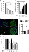
Figure 1
Figure 1
IFNγ protects GalC+ oligodendrocytes from TNFα-induced cell death and inhibits caspase activation. (a) Oligodendrocyte differentiation was induced with thyroid hormone for 5–7 days and treated with TNFα (0.1, 1, 10 and 100 ng/ml) for 72 h. Live oligodendrocytes were MTT+ and GalC+, and had DAPI+ nuclei. Data are plotted as mean±S.E.M. of at least three independent experiments. ***P<0.0001, relative to control treatment; ANOVA followed by Bonferroni's post hoc test. (b) Oligodendrocyte viability was determined after 72 h in the presence of 20 ng/ml TNFα and increasing concentrations of IFNγ (0.1, 1, 10 and 100 ng/ml). **P<0.001, relative to control treatment; ANOVA followed by Bonferroni's post hoc test. (c) Oligodendrocytes were treated with IFNγ and/or with TNFα for 72 h and immunostained with anti-activated caspase-3 antibody, anti-GalC and DAPI. Note the pyknotic nucleus in the TNFα condition. Images were optimized for brightness and contrast. Scale bars, 25 μm. (d) Oligodendrocytes treated as in b were quantified and expressed as % activated caspase-3+/GalC+ cells. Data are plotted as mean±S.E.M. of at least three independent experiments. **P<0.001 relative to control treatment; ANOVA followed by Bonferroni's post hoc test. (e) IFNγ promoted oligodendrocyte protection from TNFα while control conditioned media (Ctl-CM), IFNγ-treated oligodendrocyte CM (IFNγ-CM) and IFNγ blocking antibodies plus IFNγ failed to protect oligodendrocytes. Data are plotted as mean±S.E.M. of at least three independent experiments. ***P<0.0001, **P<0.001 relative to control treatment; ANOVA followed by Bonferroni's post hoc test. NS, no significant difference
Figure 2
Figure 2
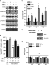
IFNγ activates Jak/STAT signaling in oligodendrocytes. (a) Whole-cell lysates were obtained from oligodendrocytes treated as indicated and probed by western blotting for pSTAT1 (Y701), STAT1 and STAT3, normalized to β-tubulin. pSTAT1 (Y701) levels were represented as pSTAT1 (Y701)/STAT1. (bd) Oligodendrocytes were treated as indicated, and RNA was isolated for RT-qPCR of IRF-1, SOCS1, SOCS3 and GAPDH. Data are plotted as mean±S.E.M. of at least three independent experiments. ***P<0.0001, **P<0.001 compared with control treatment; ANOVA followed by Bonferroni's post hoc test
Figure 3
Figure 3
Jak activation is required for oligodendrocyte protection. (a) Dose–response curve of Jak inhibitor AG-490 (0.1, 1, 5, 10 and 100 μM) on oligodendrocytes after 3 days in culture. Data are plotted as mean±S.E.M. of at least three independent experiments. ***P<0.0001 relative to DMSO vehicle control; ANOVA followed by Bonferroni's post hoc test. (b) AG-490 (1 μM) effectively inhibited STAT1 phosphorylation. Whole-cell lysates were obtained from oligodendrocytes treated with AG-490 or DMSO vehicle control and probed with antibodies against phospho-STAT1 (Y701) and β-tubulin. (c) Oligodendrocytes were treated with IFNγ and/or TNFα in the presence of AG-490 (1 μM) or DMSO vehicle control for 72 h. Data are plotted as mean±S.E.M. of technical triplicates from a representative experiment repeated three times. ***P<0.0001, **P<0.01 relative to DMSO vehicle control; ANOVA followed by Bonferroni's post hoc test. (d) Oligodendrocytes were infected with lentivirus (pLKO.1) expressing either scrambled shRNA or STAT1-targeting shRNA for 4 h, allowed 48 h for recovery and treated with IFNγ (1, 10 and 100 ng/ml) and 20 ng/ml TNFα as indicated for 72 h. Data are plotted as mean±S.E.M. of at least three independent experiments and were compared with scrambled shRNA control. (e) Oligodendrocytes were infected with lentivirus (pLKO.1) expressing either scrambled shRNA or STAT1-targeting shRNA for 4 h, allowed 48 h for recovery and whole-cell lysates were collected for western blotting to determine the degree of STAT1 knockdown. Lysates were probed with antibodies against STAT1 and β-tubulin. Lentiviral shRNA constructs targeting STAT1 protein specifically reduced STAT1 expression by >90% compared with scrambled control shRNA. (f) Oligodendrocytes were infected with lentivirus expressing scrambled shRNA as in e and treated as indicated. Data are plotted as mean±S.E.M. of at least three independent experiments. *P<0.05 compared with control treatment by ANOVA followed by Bonferroni's post hoc test. NS, not significant
Figure 4
Figure 4
PKR activation is required for IFNγ–mediated oligodendrocyte protection. (a) Whole-cell lysates were obtained from oligodendrocytes treated as indicated for 72 h and probed by western blotting for PKR, phospho-eIF2α, eIF2α, phospho-PERK, PERK and β-tubulin. (b) Oligodendrocytes were treated as indicated, and RNA was isolated for RT-qPCR of Chop and GAPDH. Data are plotted as mean±S.E.M. of at least three independent experiments. *P<0.05, **P<0.01 compared with control treatment; ANOVA followed by Bonferroni's post hoc test. (c) Oligodendrocytes were infected with lentivirus expressing either scrambled shRNA or (pGIPZ) PKR-targeting shRNA for 4 h, allowed 48 h for recovery and whole-cell lysates were collected for western blotting to determine the degree of PKR knockdown. Lysates were probed with antibodies against PKR and β-tubulin. Lentiviral shRNA constructs targeting PKR protein specifically reduced STAT1 expression by >80% compared with scrambled control shRNA. (d) Oligodendrocytes were infected with lentivirus expressing scrambled shRNA (see also Figure 3d for full set of scrambled shRNA controls) or (pGIPZ) PKR-targeting shRNA for 4 h, allowed 48 h for recovery and treated with IFNγ and/or TNFα. ***P<0.0001 compared with scrambled shRNA-infected, control-treated oligodendrocytes; ANOVA followed by Bonferroni's post hoc test. (e) RNA was isolated as in b and levels of BiP mRNA were normalized to that of GAPDH
Figure 5
Figure 5
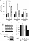
IFNγ-mediated protection requires activation of NF-κB. (a) Oligodendrocytes were infected with lentivirus containing NF-κB luciferase reporter constructs for 16 h and allowed 48 h for recovery. Treatment was as indicated for 3, 24 or 48 h. Data are plotted as mean±S.E.M. of at least three independent experiments. *P<0.05, compared with control treatment; ANOVA followed by Bonferroni's post hoc test. (b) Whole-cell lysates were obtained from oligodendrocytes treated for 24 h as indicated and probed by western blotting for pIκB-α (S32/36), IκB-α and β-actin. Protein expression was quantified and normalized to actin intensity. (c) Oligodendrocytes were infected with lentivirus (pCDH) containing either no insert (empty vector; pCDH-e.v.) or cDNA encoding FLAG-tagged, non-phosphorylatable IκB (pCDH-FLAG-IκB(SR) for 16 h, allowed 48 h for recovery and whole-cell lysates were collected to determine the expression of transgene. Lysates were probed with antibodies against FLAG and β-tubulin. (d) Oligodendrocytes were infected with lentivirus (pCDH) as in c and treated as indicated. Data are plotted as mean±S.E.M. of at least three independent experiments. **P<0.0001 compared with e.v.-infected, control-treated oligodendrocytes; ANOVA followed by Bonferroni's post hoc test
Figure 6
Figure 6
IFNγ significantly increases cFLIPL in a Jak-dependent manner, which is both sufficient and necessary for protection from TNFα. (a) Oligodendrocytes were treated as indicated and RNA was isolated for RT-qPCR of cFLIP and GAPDH. Data are plotted as mean±S.E.M. of at least three independent experiments. *P<0.05, **P<0.01 compared with control treatment; ANOVA followed by Bonferroni's post hoc test. (b) Whole-cell lysates were obtained from oligodendrocytes treated for 72 h as indicated and probed by western blotting for cFLIP, activated caspase-8 and β-tubulin. Protein expression was quantified and normalized to tubulin intensity. (c) Oligodendrocytes were infected with control (RFP) or cFLIP overexpressing lentivirus (pCDH) constructs for 16 h, given 48 h to recover and treated with TNFα for 72 h. Cells were then fixed and stained with anti-GalC and cFLIP antibodies. Images show representative cell cultures treated with the indicated conditions. Images were optimized for brightness and contrast. Note the pyknotic nucleus in the control virus, TNFα condition (c′). (d) Oligodendrocytes were infected with lentivirus (pLKO.1) expressing either scrambled shRNA or cFLIP-targeting shRNA for 16 h, allowed 48 h for recovery and whole-cell lysates were collected to determine the degree of cFLIP knockdown. Lysates were probed with antibodies against cFLIP and β-tubulin. Lentiviral shRNA constructs targeting cFLIP protein specifically reduced cFLIP expression by >50% compared with scrambled control shRNA. (e) Oligodendrocytes were infected with lentivirus (pLKO.1) expressing either scrambled shRNA or cFLIP-targeting shRNA for 16 h, allowed 48 h for recovery and treated for 72 h with IFNγ and/or TNFα. (A full set of scr ShRNA control vectors is shown in Figure 3f). Data are plotted as mean±S.E.M. of at least three independent experiments. *P<0.05, **P<0.01 compared with scrambled shRNA-infected, control-treated oligodendrocytes; ANOVA followed by Bonferroni's post hoc test. (f) Oligodendrocytes were pre-treated with 5 μm AG-490 or DMSO for 16 h, then co-treated with DMSO, 5 μm AG-490, IFNγ and/or TNFα as indicated for 6 h. RNA was isolated for RT-qPCR of cFLIP and GAPDH. Data are plotted as mean±S.E.M. of three independent experiments. *P<0.05, ***P<0.001; ANOVA followed by Bonferroni's post hoc test
Figure 7
Figure 7
cFLIP knockdown during sustained neuroinflammation in vivo increases oligodendrocyte death and decreases myelin staining intensity. (a and b) Adult IL-1βXAT mice were injected with transgene activating Cre-expressing lentivirus and either scrambled shRNA (Cre-virus scr-shRNA) or cFLIP-targeting (Cre-virus cFLIP) shRNA and aged for 2 weeks. Brain sections were stained with GSTπ antibodies, DAPI and for TUNEL. (b′) Higher magnification views of TUNEL+/GSTπ+ oligodendrocytes. (c) Quantification of TUNEL+/GSTπ+ oligodendrocytes per field. Data are plotted as mean±S.D. of five fields per section from two mice. *P<0.05, t-test. (d) Adult IL-1βXAT mice were injected with Cre-expressing virus or control virus and RNA was isolated from brain for RT-PCR of TNFα, IFNγ and cFLIP. Note that the upregulation of cFLIP levels during IL-1β-mediated inflammation coincides with the absence of oligodendrocytes death in this mouse model. (e) Sections from adult IL-1βXAT mice injected with the Cre-expressing virus into the corpus callosum of both hemispheres, with one side also receiving a cFLIP-specific shRNA-expressing virus (right, r) and the contralateral side receiving a non-targeting, scrambled shRNA-expressing virus (left, l) that were stained with FluoroMyelin. (f) Quantification of fluorescence intensity in arbitrary units from three sections per mouse and two mice. *P<0.05 t-test

Similar articles

Cited by

References

    1. Agresti C, D'Urso D, Levi G. Reversible inhibitory effects of interferon-gamma and tumour necrosis factor-alpha on oligodendroglial lineage cell proliferation and differentiation in vitro. Eur J Neurosci. 1996;8:1106–1116. - PubMed
    1. Tanner DC, Cherry JD, Mayer-Proschel M. Oligodendrocyte progenitors reversibly exit the cell cycle and give rise to astrocytes in response to interferon-gamma. J Neurosci. 2011;31:6235–6246. - PMC - PubMed
    1. Andrews T, Zhang P, Bhat NR. TNFalpha potentiates IFNgamma-induced cell death in oligodendrocyte progenitors. J Neurosci Res. 1998;54:574–583. - PubMed
    1. Baerwald KD, Popko B. Developing and mature oligodendrocytes respond differently to the immune cytokine interferon-gamma. J Neurosci Res. 1998;52:230–239. - PubMed
    1. Lin W, Harding HP, Ron D, Popko B. Endoplasmic reticulum stress modulates the response of myelinating oligodendrocytes to the immune cytokine interferon-gamma. J Cell Biol. 2005;169:603–612. - PMC - PubMed

Publication types

MeSH terms