. 2015 Jan 29;6(1):e1621.
doi: 10.1038/cddis.2014.591.

Targeting MKK3 as a novel anticancer strategy: molecular mechanisms and therapeutical implications

Affiliations

Targeting MKK3 as a novel anticancer strategy: molecular mechanisms and therapeutical implications

S Baldari et al. Cell Death Dis. .

Abstract

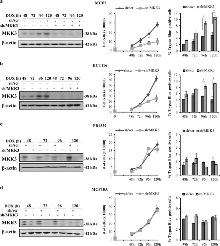
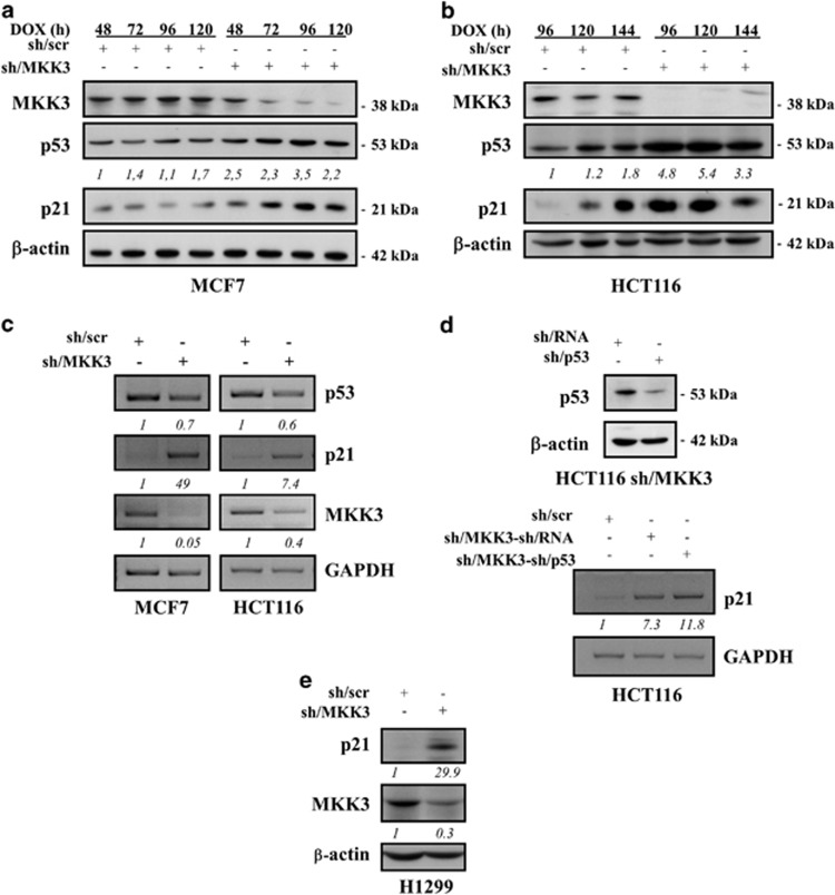
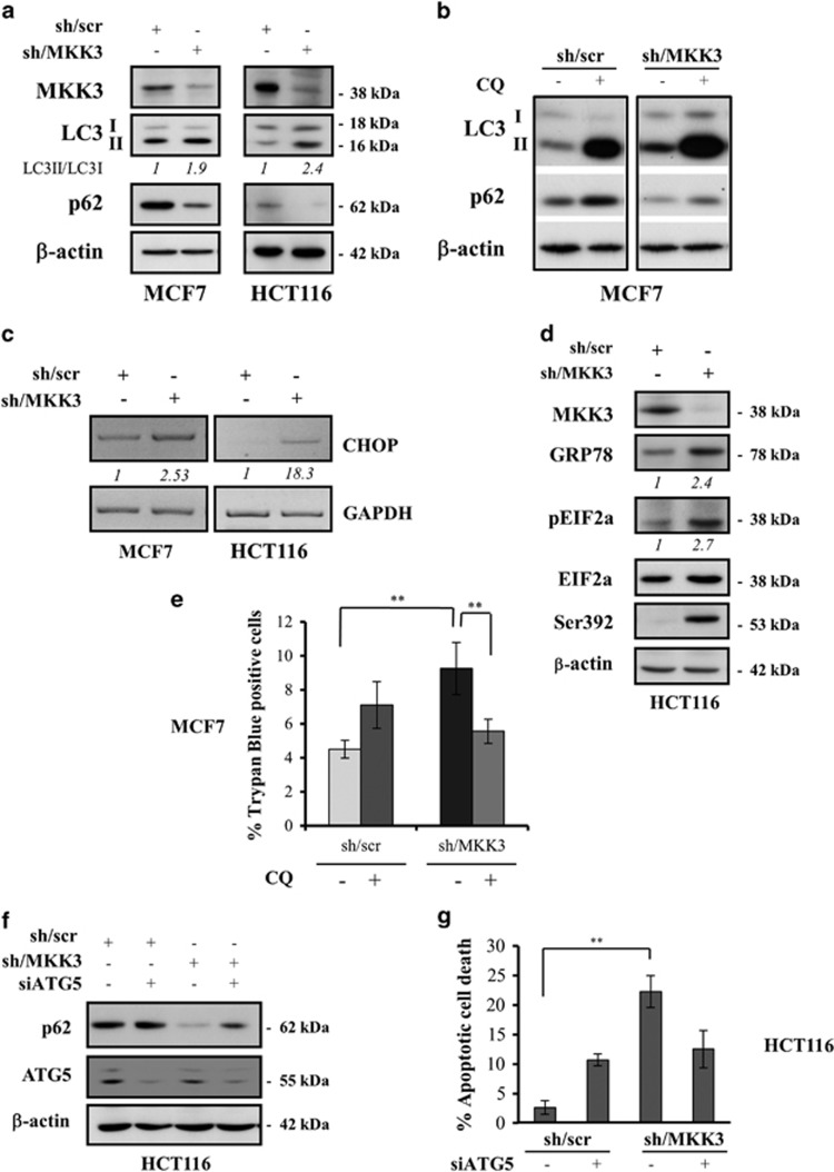
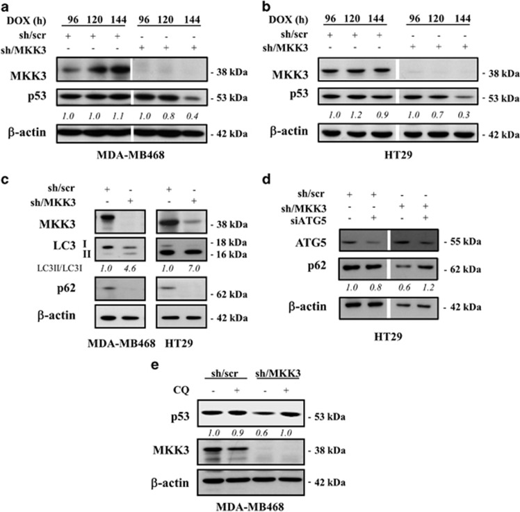
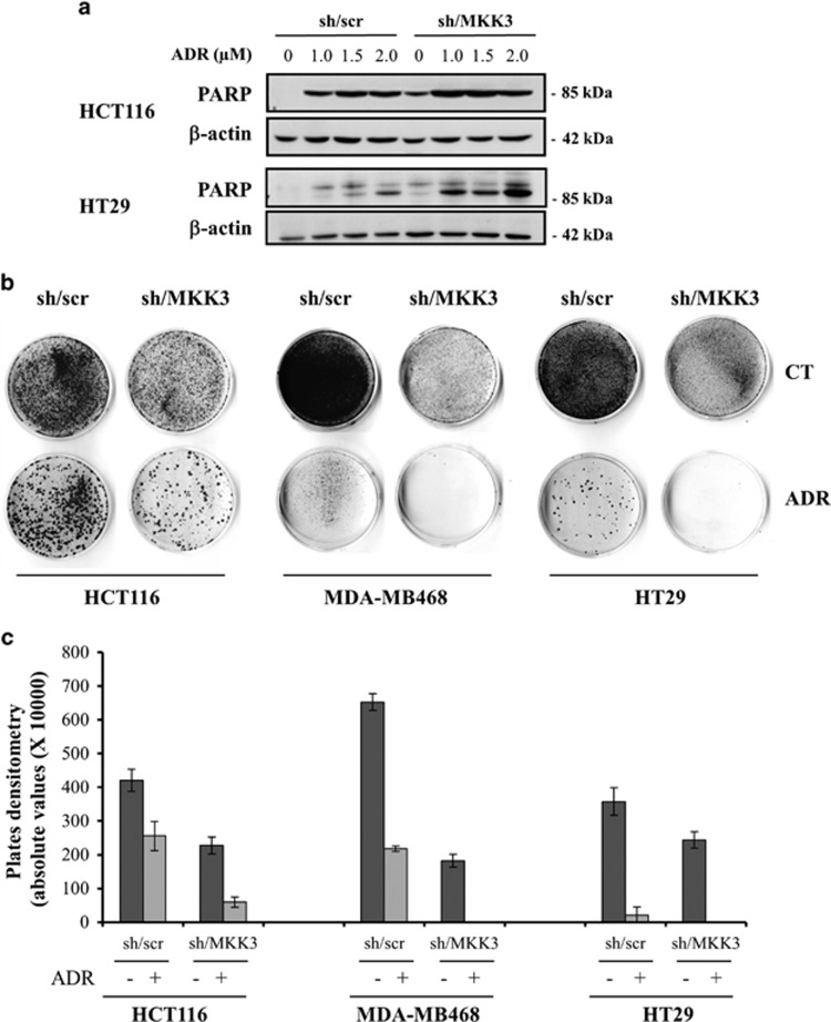
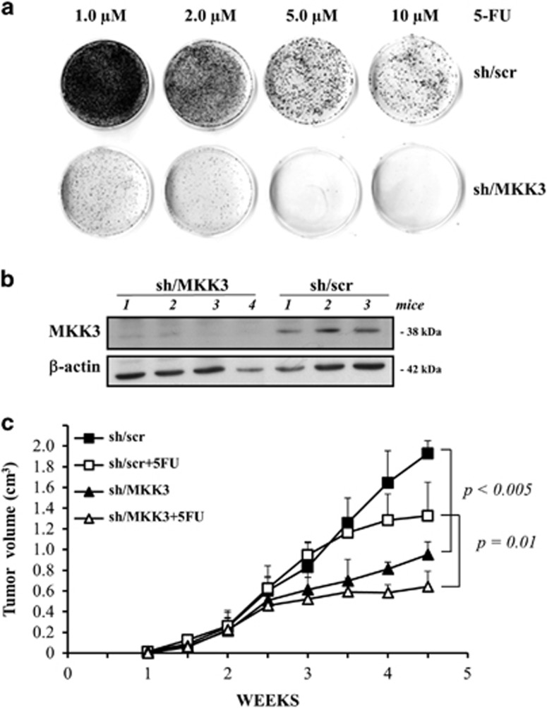
Mitogen-activated protein kinase kinase 3 (MAP2K3, MKK3) is a member of the dual specificity protein kinase group that belongs to the MAP kinase kinase family. This kinase is activated by mitogenic or stress-inducing stimuli and participates in the MAP kinase-mediated signaling cascade, leading to cell proliferation and survival. Several studies highlighted a critical role for MKK3 in tumor progression and invasion, and we previously identified MKK3 as transcriptional target of mutant (mut) p53 to sustain cell proliferation and survival, thus rendering MKK3 a promising target for anticancer therapies. Here, we found that targeting MKK3 with RNA interference, in both wild-type (wt) and mutp53-carrying cells, induced endoplasmic reticulum stress and autophagy that, respectively, contributed to stabilize wtp53 and degrade mutp53. MKK3 depletion reduced cancer cell proliferation and viability, whereas no significant effects were observed in normal cellular context. Noteworthy, MKK3 depletion in combination with chemotherapeutic agents increased tumor cell response to the drugs, in both wtp53 and mutp53 cancer cells, as demonstrated by enhanced poly (ADP-ribose) polymerase cleavage and reduced clonogenic ability in vitro. In addition, MKK3 depletion reduced tumor growth and improved biological response to chemotherapeutic in vivo. The overall results indicate MKK3 as a novel promising molecular target for the development of more efficient anticancer treatments in both wtp53- and mutp53-carrying tumors.


Figures

Figure 1
Figure 1
MKK3 depletion is detrimental to cancer but not normal cell proliferation and survival. Efficient MKK3 depletion was achieved in all tested lines (ad, left panels): Engineered MCF7 (a), HCT116 (b), FB1329 (c) MCF10A (d) -sh/scr and -sh/MKK3 sublines (1.5 × 10⁵ cells/60 mm dish) were challenged with DOX (1.0 μg/ml) and cells collected time dependently (48, 72, 96 and 120 h) to assess the MKK3 depletion efficiency. Then, protein lysates (30 μg /lane) were resolved in SDS–polyacrylamide gel electrophoresis and filter analyzed by western blot analysis with specific anti-MKK3 and anti-β-actin (loading control) antibodies. MKK3 depletion affects cell proliferation and viability of wtp53 tumor but not normal cells (a–d, right panels). Engineered MCF7 (a), HCT116 (b), FB1329 (c) MCF10A (d) -sh/scr and -sh/MKK3 sublines were seeded (2 × 10⁴ cells/6-well plates) in DOX conditions. At the indicated time points, cells were harvested and quantified for viable and death cells by Trypan blue exclusion assays. Results are reported as means and S.D. of three independent experiments. *The significance (P<0.05) of death percent in sh/MKK3 with respect to sh/scr subline
Figure 2
Figure 2
MKK3 depletion stabilizes wtp53 protein. Engineered MCF7 (a) and HCT116 (b) -sh/scr and -sh/MKK3 sublines were cultured with DOX (1.0 μg/ml) and collected at the indicated time points. Protein lysates (30 μg/lane) were analyzed by western blot analysis with anti-MKK3-, anti-p53-, anti-p21-, and anti-β-actin (loading control)-specific antibodies. Densitometry was performed with ImageJ software and relative p53 band intensity normalized to β-actin and quantified with respect to controls (sh-scr) set to 1.0. (c) Semi-quantitative RT-PCR was performed on total RNAs isolated from engineered MCF7 (left panel) and HCT116 (right panel) -sh/scr and sh/MKK3 sublines maintained 72 h with DOX. PCR was performed with specific set of primers. GAPDH was used as housekeeping gene. RT-PCR images were acquired by Bio-Rad Universal Hood II gel-imager. Densitometry was performed with ImageJ software and relative p53, p21, and MKK3 band intensity normalized to GAPDH and quantified with respect to controls (sh-scr) set to 1.0. (d) Upper panel: HCT116-sh/MKK3 sublines were transfected with sh/p53 or control sh/RNA carrying vector and efficiency of p53 depletion detected by western blotting (30 μg/lane). Lower panel: HCT116-sh/scr, -sh/MKK3-sh/RNA, and -sh/MKK3-sh/p53 were cultured 72 h with DOX, then total RNAs were isolated and semi-quantitative RT-PCR performed with set of primers specific to p21 and GAPDH (housekeeping gene). Images were acquired by Bio-Rad Universal Hood II gel-imager, and densitometry performed with ImageJ software. Relative p21 band intensity was normalized to GAPDH and quantified with respect to controls (sh-scr) set to 1.0. (e) Engineered H1299-sh/scr and -sh/MKK3 sublines were cultured with DOX (1.0 μg/ml) and collected 96 h later. Protein lysates (30 μg/lane) were analyzed by western blot analysis with anti-MKK3-, anti-p21- and anti-β-actin (loading control)-specific antibodies. Densitometry was performed with ImageJ software and relative p21 and MKK3 band intensity normalized to β-actin and quantified with respect to controls (sh-scr) set to 1.0
Figure 3
Figure 3
MKK3 depletion induces autophagic cell death and ER stress in wtp53 cancer cells. Markers of autophagy LC3-I, LC3-II, and p62 upon MKK3 depletion were assessed by western blotting (a) in wtp53 MCF7 (left panel) and HCT116 (right panel) engineered -sh/scr and sh/MKK3 sublines. Seeded cells (1.5 × 10⁵ cells/60 mm dish) were collected at 72 h post DOX delivery, and protein lysates (15 μg /lane) resolved in SDS–polyacrylamide gel electrophoresis (PAGE) and probed with anti-MKK3-, anti-LC3-, and anti-SQSTM1/p62-specific antibodies. Densitometry analyses were performed with ImageJ software and LC3-II band intensity normalized to β-actin and quantified with respect to control tumors (sh/scr) set to 1.0. (b) MCF7-sh/scr and -sh/MKK3 sublines cultured in DOX condition (72 h) in the presence/absence of 25 μM CQ (48 h), then protein lysate were analyzed by western blot analysis with antibodies specific to LC3, SQSTM1/p62, and β actin (loading control). (c) Engineered sh/MKK3 and sh/scr MCF7 and HCT116 cultured 48 h with DOX were collected and total RNAs analyzed by RT-PCR with set of primers specific to CHOP and GAPDH (housekeeping gene). Densitometry was performed with the ImageJ software and relative CHOP mRNA levels were normalized to GAPDH and quantified with respect to control tumors (sh-scr) set to 1.0. (d) Engineered sh/scr and sh/MKK3 HCT116 cell lines were maintained in DOX condition for 36 h, then cells were collected and protein lysates (30 μg/lane) analyzed by western blot for the presence of ER stress proteins: phosphorylated EIF2A protein was evaluated using phospho-specific antibodies. Total amount of EIF2A was determined using anti-EIF2A antibody. GRP78/Bip was also used as marker of ER stress. Ser392 was analyzed as p53 stabilization marker. β-Actin was used as loading control. Densitometry was performed with the ImageJ software and relative ER stress marker protein levels were normalized to actin and quantified with respect to control tumors (sh-scr) set to 1.0. (e) Autophagy inhibition rescues the cell death induced by MKK3 depletion. Engineered MCF7-sh/scr and -sh/MKK3 sublines were seeded (1.5 × 10⁵ cells/60 mm dish) in DOX condition for 24 h then treated/untreated with CQ (25 μM) and collected after 96 h of total DOX induction. Cell viability was evaluated with trypan blue exclusion assay. Results are reported as mean±S.D. of three independent experiments. Significance was assessed by Student's t-test, **P<0.01. (f) Engineered HCT116-sh/scr and -sh/MKK3 sublines were seeded (1.5 × 10⁵ cells/60 mm dish) and maintained in DOX condition for 48 h, then transfected with siRNA for ATG5 (si-ATG5) or with control siRNA (si-ctr). Forty-eight hours after transfection, cells were collected and protein lysates (15 μg/lane) resolved in SDS-PAGE and probed with anti-ATG5- and anti-SQSTM1/p62-specific antibodies. β-Actin was used as loading control. (g) Engineered HCT116-sh/scr and -sh/MKK3 sublines, treated as in f, were collected after 96 h of DOX induction and then stained with DAPI and ethidium bromide and analyzed by fluorescent microscope. The number of apoptotic dead cells was calculated and reported as a percentage of the total number of cell counted. Results are reported as means and S.D. of three independent experiments. **The significance (P<0.01) of death percent in sh/MKK3 with respect to sh/scr subline
Figure 4
Figure 4
MKK3 depletion reduces mutp53 protein levels through autophagy. Engineered MDA-MB468 (a) and HT29 (b) -sh/scr and -sh/MKK3 sublines were cultured with DOX (1.0 μg/ml) and collected at indicated time points. Protein lysates (30 μg/lane) were analyzed by western blot analysis with anti-MKK3-, anti-p53-, and anti-β-actin (loading control)-specific antibodies. Densitometry was performed with ImageJ software and relative p53 band intensity normalized to β-actin and quantified with respect to controls (sh-scr) set to 1.0. (c) Markers of autophagy LC3-I, LC3-II, and p62 upon MKK3 depletion were assessed by western blotting in mutp53 MDA-MB468 (left panel) and HT29 (right panel) engineered -sh/scr and sh/MKK3 sublines. Seeded cells (1.5 × 10⁵ cells/60 mm dish) were collected at 120 h post DOX delivery, and protein lysates (15 μg /lane) resolved in SDS–polyacrylamide gel electrophoresis (PAGE) and probed with anti-MKK3-, anti-LC3-, and anti-SQSTM1/p62-specific antibodies. Densitometry analyses were performed with ImageJ software and LC3-II band intensity normalized to β-actin and quantified with respect to control tumors (sh/scr) set to 1.0. (d) Engineered HT29-sh/scr and -sh/MKK3 sublines were seeded (1.5 × 10⁵ cells/60 mm dish) and maintained in DOX condition for 72 h, then transfected with siRNA for ATG5 (si-ATG5) or with control siRNA (si-ctr). Forty-eight hours after transfection, cells were collected and protein lysates (15 μg /lane) resolved in SDS-PAGE and probed with anti-ATG5- and anti-SQSTM1/p62-specific antibodies. β-Actin was used as loading control. Densitometry analyses were performed with ImageJ software and anti-SQSTM1/p62 band intensity normalized to β-actin and quantified with respect to control tumors (sh/scr) set to 1.0. (e) MDA-MB468 sh/scr and sh/MKK3 sublines were cultured 120 h with DOX and, in the last 48 h, treated/untreated with CQ (25 μM). Afterwards, cells were collected and lysate analyzed by western blot analysis with anti-p53-, anti-MKK3-, and anti-β actin (loading control)-specific antibodies
Figure 5
Figure 5
MKK3 depletion increases chemotherapeutic response in both wtp53 and mutp53 cancer cells. (a) Engineered sh/scr and sh/MKK3 HCT116 and HT29 cell lines were maintained in DOX condition for 48 h and treated/untreated for 24 h with ADR (1, 1.5, and 2 μM). Cells were then collected and protein lysates were analyzed by western blot analysis. The membrane was probed with specific anti-PARP and anti-β-actin (as loading control) antibodies. (b) Clonogenic survival assay of: HCT116 (left panel), MDA-MB468 (middle panel), and HT29 (right panel) sh/scr and sh/MKK3 cell lines, DOX-induced for 48 h and then treated with ADR 0.1 μM (HCT116 and MDA-MB468) or 0.5 μM (HT29) for 24 h. After treatment, the culture medium was replenished, and cells were maintained at 37 °C for 14 days. Grown colonies were stained with crystal violet. (c) Densitometric analyses of clonogenic assays as described in b. Plates were scanned and quantified by the ImageJ software. The graph shows the absolute values of plates densitometry. Plating was performed in triplicate
Figure 6
Figure 6
MKK3 depletion affects xenograft tumor growth and increases chemotherapeutic response in vivo. (a) Dose-response effect of 5-FU on clonogenic survival in HT29-sh/scr and sh/MKK3 cell lines, DOXI-induced for 48 h, and then treated with 5-FU (1, 2, 5, and 10 μM) for 24 h. After treatment, the culture medium was replenished, and cells were maintained at 37 °C for 14 days. Grown colonies were stained with crystal violet. (b) Efficient MKK3 depletion was achieved in vivo, assessed by western blot analysis performed on a representative number of xenograft tumors generated with sh/scr and sh/MKK3 engineered HT29 cancer cells injected in nude mice (CD1/SWISS). Numbers identify single animal. (c) After tumor nodule formation, DOX (2.0 g/l, tap water) was delivered to all mice. To assess chemotherapeutic response in MKK3-depleted tumors, 5-FU (50 mg/kg, intraperitoneal) was delivered to a subgroup of sh/scr and sh/MKK3 tumor-bearing mice (8 mice/group). Tumor growth was followed by calliper measurements twice a week. Representative data of two independent experiments are reported. Student's t-test analyses were performed to assess significance between sh/scr and sh/MKK3 tumor-bearing mice (P<0.05) and along with 5-FU treatments (P=0.01)

References

    1. Johnson GL, Lapadat R. Mitogen-activated protein kinase pathways mediated by ERK, JNK, and p38 protein kinases. Science 2002; 298: 1911–1912. - PubMed
    1. Raingeaud J, Whitmarsh AJ, Barrett T, Derijard B, Davis RJ. MKK3- and MKK6-regulated gene expression is mediated by the p38 mitogen-activated protein kinase signal transduction pathway. Mol Cell Biol 1996; 16: 1247–1255. - PMC - PubMed
    1. Han J, Lee JD, Jiang Y, Li Z, Feng L, Ulevitch RJ et al. Characterization of the structure and function of a novel MAP kinase kinase (MKK6). J Biol Chem 1996; 271: 2886–2891. - PubMed
    1. Raingeaud J, Gupta S, Rogers JS, Dickens M, Han J, Ulevitch RJ et al. Pro-inflammatory cytokines and environmental stress cause p38 mitogen-activated protein kinase activation by dual phosphorylation on tyrosine and threonine. J Biol Chem 1995; 270: 7420–7426. - PubMed
    1. Pearson G, Robinson F, Beers Gibson T, Xu BE, Karandikar M, Berman K et al. Mitogen-activated protein (MAP) kinase pathways: regulation and physiological functions. Endocr Rev 2001; 22: 153–183. - PubMed

Publication types

MeSH terms