. 2015 Jan 29;6(1):e1624.
doi: 10.1038/cddis.2014.546.

DRAM1 regulates apoptosis through increasing protein levels and lysosomal localization of BAX

Affiliations

DRAM1 regulates apoptosis through increasing protein levels and lysosomal localization of BAX

J-J Guan et al. Cell Death Dis. .

Abstract

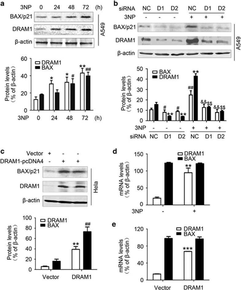
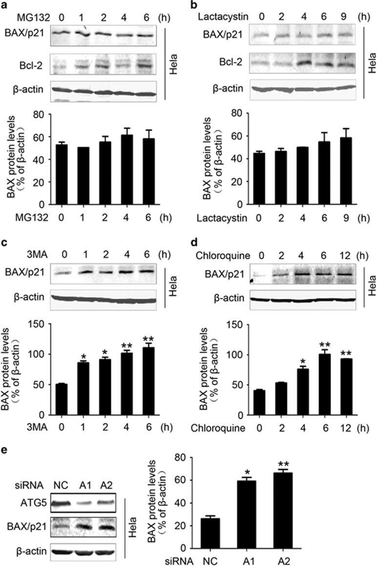
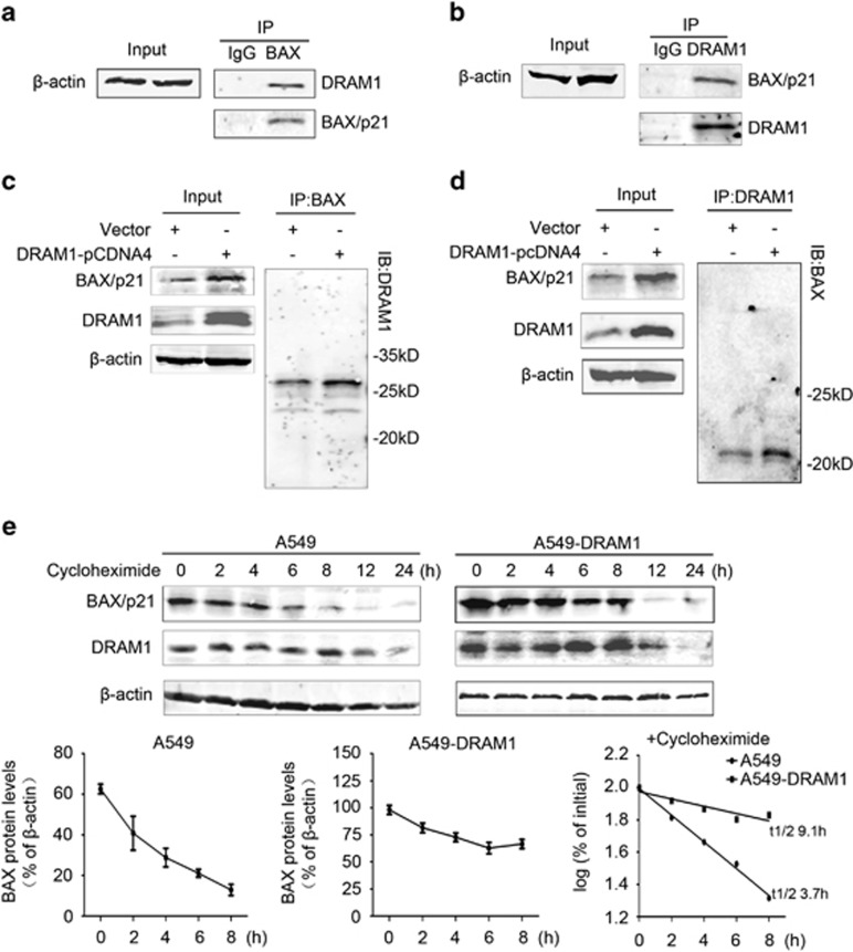
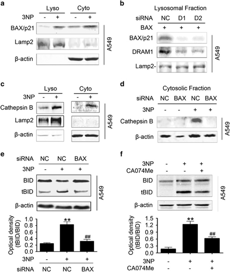
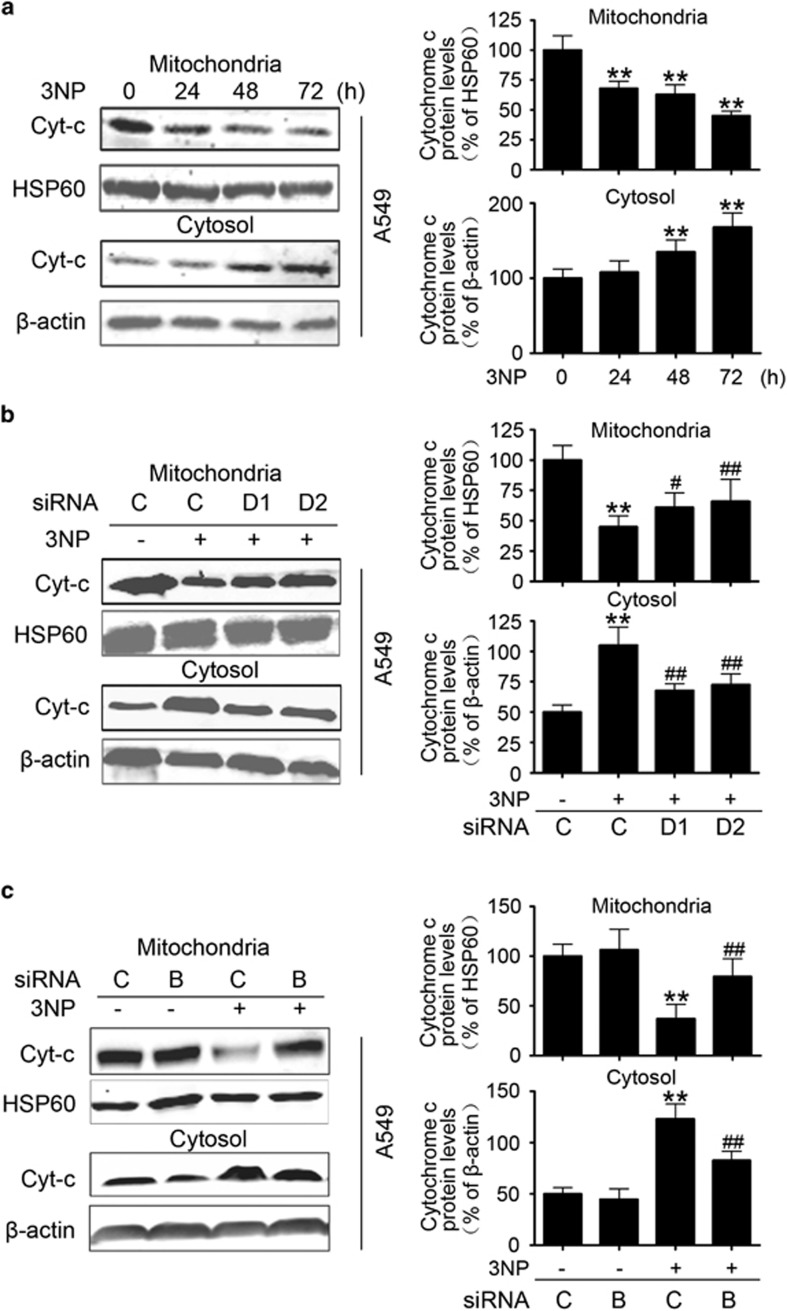
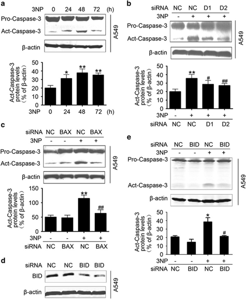
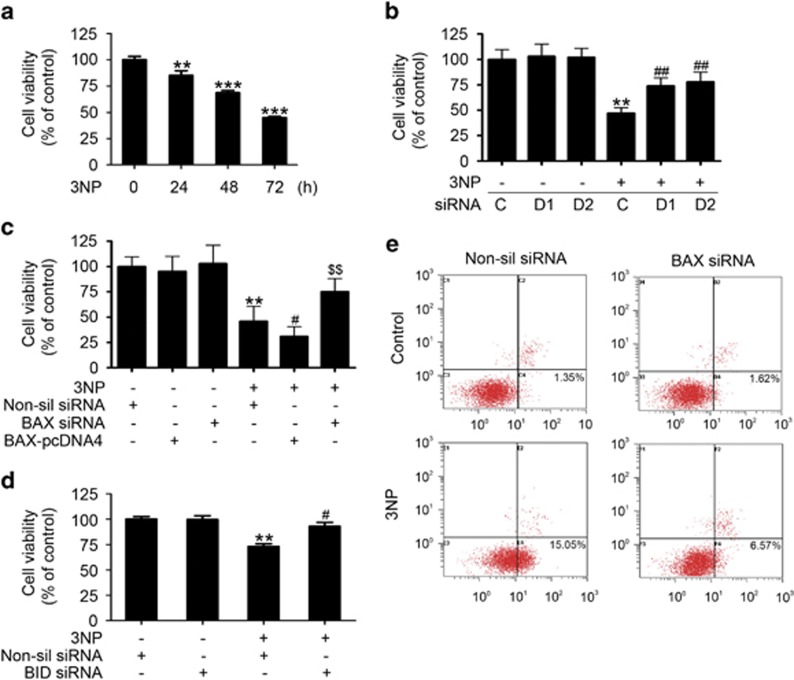
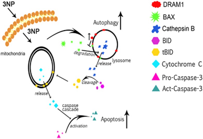
DRAM1 (DNA damage-regulated autophagy modulator 1) is a TP53 target gene that modulates autophagy and apoptosis. We previously found that DRAM1 increased autophagy flux by promoting lysosomal acidification and protease activation. However, the molecular mechanisms by which DRAM1 regulates apoptosis are not clearly defined. Here we report a novel pathway by which DRAM1 regulates apoptosis involving BAX and lysosomes. A549 or HeLa cells were treated with the mitochondrial complex II inhibitor, 3-nitropropionic acid (3NP), or an anticancer drug, doxorubicin. Changes in the protein and mRNA levels of BAX and DRAM1 and the role of DRAM1 in BAX induction were determined. The interaction between DRAM1 and BAX and its effect on BAX degradation, BAX lysosomal localization, the release of cathepsin B and cytochrome c by BAX and the role of BAX in 3NP- or doxorubicin-induced cell death were studied. The results showed that BAX, a proapoptotic protein, was induced by DRAM1 in a transcription-independent manner. BAX was degraded by autophagy under basal conditions; however, its degradation was inhibited when DRAM1 expression was induced. There was a protein interaction between DRAM1 and BAX and this interaction prolonged the half-life of BAX. Furthermore, upregulated DRAM1 recruited BAX to lysosomes, leading to the release of lysosomal cathepsin B and cleavage of BID (BH3-interacting domain death agonist). BAX mediated the release of mitochondrial cytochrome c, activation of caspase-3 and cell death partially through the lysosome-cathepsin B-tBid pathway. These results indicate that DRAM1 regulates apoptosis by inhibiting BAX degradation. In addition to mitochondria, lysosomes may also be involved in BAX-initiated apoptosis.


Figures

Figure 1
Figure 1
DRAM1 increases BAX protein levels independent of transcription. (a) A549 cells were treated with 3NP (500 μM) and harvested 24, 48 and 72 h later. Bars represent mean±S.E.; n=4; **P<0.01 versus 0 h; ##P<0.01 versus 0 h. (b) A549 cells were transfected with DRAM1 small interfering RNA (siRNA) or a non-silencing siRNA for 24 h. Cells were then treated with or without 3NP (500 μM) for another 24 h. Bars represent mean±SE; n=4; **P<0.01 versus NC; #P<0.05; ##P<0.01 versus NC; $$P<0.01 versus NC+3NP; &&P<0.01 versus NC+3NP. (c) HeLa cells were transfected with a vector or DRAM1 plasmid for 48 h. Bars represent mean±S.E.; n=4; **P<0.01 versus vector; ##P<0.01 versus vector. (d) HeLa cells were treated with 3NP (500 μM) for 48 h. RNAs were isolated and amplified with qRT-PCR. Bars represent mean±S.E.; n=4; **P<0.01 versus control. (e) HeLa cells were transfected with a vector or DRAM1 plasmid for 48 h. RNAs were isolated and amplified with qRT-PCR. Bars represent mean±S.E.; n=4; ***P<0.001 versus vector. NC, negative control
Figure 2
Figure 2
BAX is degraded by autophagy and DRAM1 interferes with BAX degradation. (a and b) Cells were treated with UPS inhibitor MG132 (40 μM) for 1, 2, 4 and 6 h or lactacystin (4 μM) for 2, 4, 6, and 9 h. The cell lysates were subjected to immunoblotting. β-Actin served as a loading control and Bcl-2 served as a positive control. Bars represent mean±S.E.; n=3. (c) HeLa cells were treated with an autophagy inhibitor 3MA (1.5 mg/ml) for 1, 2, 4 and 6 h. Bars represent mean±S.E.; n=4; *P<0.05; **P<0.01 versus 0 h. (d) HeLa cells were treated with an autophagy inhibitor chloroquine (20 μM) for 2, 4, 6 and 12 h. Bars represent mean±S.E.; n=4; *P<0.05; **P<0.01 versus 0 h. (e) Cells were transfected with Atg5 small interfering RNA (siRNA) for 48 h to inhibit autophagy. Bars represent mean±S.E.; n=4; *P<0.05; **P<0.01 versus NC. NC, negative control; 3MA, 3-methyladenine
Figure 3
Figure 3
DRAM1 interacts with BAX. (a) Cell lysates were immunoprecipitated (IP) with an anti-BAX antibody and then immunoblotted (IB) against DRAM1. (b) Cell lysates were IP with an anti-DRAM1 antibody and then IB against BAX. (c) Cell lysates from cells transfected with vector or DRAM1-pcDNA4 were IP with an anti-BAX antibody and then IB against DRAM1. (d) Cell lysates as mentioned in panel c were IP with an anti-DRAM1 antibody and then IB against BAX. (e) The half-life of BAX was prolonged in DRAM1-overexpressing cells. A549 cells and those stably transfected with DRAM1 were treated with cycloheximide (50 μg/ml). Cells were harvested at 0, 2, 4, 6, 8, 12 and 24 h after treatment. Bars represent mean±S.E.; n=3. The density of immunoreactivity for each band was measured and normalized to the density at t=0 (100%). The log10 of the percentage of density was plotted versus time, and the t1/2 was calculated from the log 10 of 50%
Figure 4
Figure 4
BAX translocates to lysosome, releases cathepsin B and activates the BID cleavage. (a) A549 cells were treated with 3NP (500 μM) for 48 h. The cytoplasmic (Cyto) and lysosomal (Lyso) fractions were fractionated and then subjected to sodium dodecyl sulfate-polyacrylamide gel electrophoresis (SDS-PAGE). (b) Lysosomes from cells transfected with or without DRAM1 small interfering RNA (siRNA) were fractionated and then incubated with BAX protein at 37 °C for 2 h. The lysosomal fractions were subjected to SDS-PAGE. (c) A549 cells were treated with 3NP (500 μM) for 48 h. The Cyto and Lyso were fractionated and then subjected to SDS-PAGE. (d) A549 cells were transfected with BAX siRNA or a non-silencing siRNA for 24 h and then treated with 3NP (500 μM) for another 24 h; the cytoplasm was fractionated for western blot analysis. (e) A549 cells were transfected with BAX siRNA or a non-silencing siRNA for 24 h and then treated with or without 3NP (500 μM) for another 24 h. Bars represent mean±S.E.; n=4; **P<0.01 versus NC; ##P<0.01 versus 3NP+NC. (f) A549 cells were treated with 3NP (500 μM) and the cathepsin B inhibitor for 24 h. Bars represent mean±S.E.; n=4; **P<0.01 versus control; ##P<0.01 versus 3NP. NC, negative control
Figure 5
Figure 5
BAX mediates the proapoptotic role of DRAM1. (a) A549 cells were treated with 3NP (500 μM) and harvested 24, 48 and 72 h later for the preparation of mitochondrial and cytosolic fractions. Bars represent mean±S.E.; n=3; **P<0.01 versus 0h. (b) A549 cells were transfected with DRAM1 small interfering RNA (siRNA) or non-silencing (Non-sil) siRNA for 24 h and then treated with 3NP (500 μM) for 24 h. Bars represent mean±S.E.; n=3; **P<0.01 versus NC; #P<0.05; ##P<0.01 versus 3NP+NC. (c) A549 cells were transfected with BAX siRNA or Non-sil siRNA for 24 h and then treated with 3NP (500 μM) for 24 h. Bars represent mean±S.E.; n=3; **P<0.01 versus NC; ##P<0.01 versus 3NP+NC. NC, negative control
Figure 6
Figure 6
BAX mediates proapoptotic role of DRAM1 partially through tBID. (a) Immunoblot analysis of the activation of caspase-3. A549 cells were treated with 3NP (500 μM) for 24, 48 and 72 h; the activation of caspase-3 was determined by immunoblotting. Bars represent mean±S.E.; n=3; *P<0.05, **P<0.01 versus 0 h. (b) A549 cells were transfected with DRAM1 small interfering RNA (siRNA) or non-silencing (Non-sil) siRNA for 24 h and then treated with 3NP (500 μM) for 24 h. Bars represent mean±S.E.; n=3, **P<0.01 versus NC; #P<0.05, ##P<0.01 versus 3NP+NC. (c) A549 cells were transfected with BAX siRNA or Non-sil siRNA for 24 h and then treated with 3NP (500 μM) for 24 h. Bars represent mean±S.E.; n=3, **P<0.01 versus NC; ##P<0.01 versus 3NP+NC. (d) Efficiency of siRNA-mediated downregulation of BID. (e) A549 cells were transfected with BID siRNA or Non-sil siRNA for 24 h and then treated with 3NP (500 μM) for another 24 h. Active caspase-3 was determined by immunoblotting. Bars represent mean±S.E.; n=3; *P<0.05 versus NC, #P<0.05 versus NC+3NP. NC, negative control
Figure 7
Figure 7
DRAM1 regulates cell death through BAX. (a) A549 cells were treated with 3NP (500 μM) for 24, 48 and 72 h, and the cell viability was evaluated. Bars represent mean±S.E.; n=4; **P<0.01; ***P<0.001 versus 0 h. (b) Cell viability was evaluated after transfection of cells with DRAM1 small interfering RNA (siRNA) in the presence or absence of 3NP (500 μM). Bars represent mean±S.E.; n=4; **P<0.01 versus NC; ##P<0.01 versus NC+3NP. (c) Cell viability was evaluated after transfection of cells with BAX-pcDNA4 or BAX siRNA in the presence or absence of 3NP (500 μM). Bars represent mean±S.E.; n=4; **P<0.01 versus NC; #P<0.05 versus NC+3NP; $$P<0.01 versus NC+3NP. (d) A549 cells were transfected with BID siRNA for 24 h and then treated with 3NP (500 μM) for another 24 h. Bars represent mean±S.E.; n=3; **P<0.01 versus NC; #P<0.05 versus 3NP+non-silencing (Non-sil) siRNA. (e) A549 cells were transfected with BAX siRNA or Non-sil siRNA for 24 h and then treated with 3NP (500 μM) for another 24 h. After treatment, apoptotic cells were determined with FACS after Annexin V-FITC and PI staining. NC, Negative control. NC, negative control
Figure 8
Figure 8
Proposed model for the action of DRAM1 in 3NP- and doxorubicin-induced autophagy and apoptosis

Similar articles

Cited by

References

    1. Nezis IP, Shravage BV, Sagona AP, Johansen T, Baehrecke EH, Stenmark H. Autophagy as a trigger for cell death: autophagic degradation of inhibitor of apoptosis dBruce controls DNA fragmentation during late oogenesis in Drosophila. Autophagy 2010; 6: 1214–1215. - PMC - PubMed
    1. McPhee CK, Logan MA, Freeman MR, Baehrecke EH. Activation of autophagy during cell death requires the engulfment receptor Draper. Nature 2010; 465: 1093–1096. - PMC - PubMed
    1. Pattingre S, Tassa A, Qu X, Garuti R, Liang XH, Mizushima N et al. Bcl-2 antiapoptotic proteins inhibit Beclin 1-dependent autophagy. Cell 2005; 122: 927–939. - PubMed
    1. Crighton D, Wilkinson S, Ryan KM. DRAM links autophagy to p53 and programmed cell death. Autophagy 2007; 3: 72–74. - PubMed
    1. Crighton D, Wilkinson S, O'Prey J, Syed N, Smith P, Harrison PR et al. DRAM, a p53-induced modulator of autophagy, is critical for apoptosis. Cell 2006; 126: 121–134. - PubMed

Publication types