. 2015 Mar 1;142(5):931-42.
doi: 10.1242/dev.112904. Epub 2015 Jan 29.

Contributions of Costal 2-Fused interactions to Hedgehog signaling in Drosophila

Affiliations

Contributions of Costal 2-Fused interactions to Hedgehog signaling in Drosophila

Eva V Zadorozny et al. Development. .

Abstract

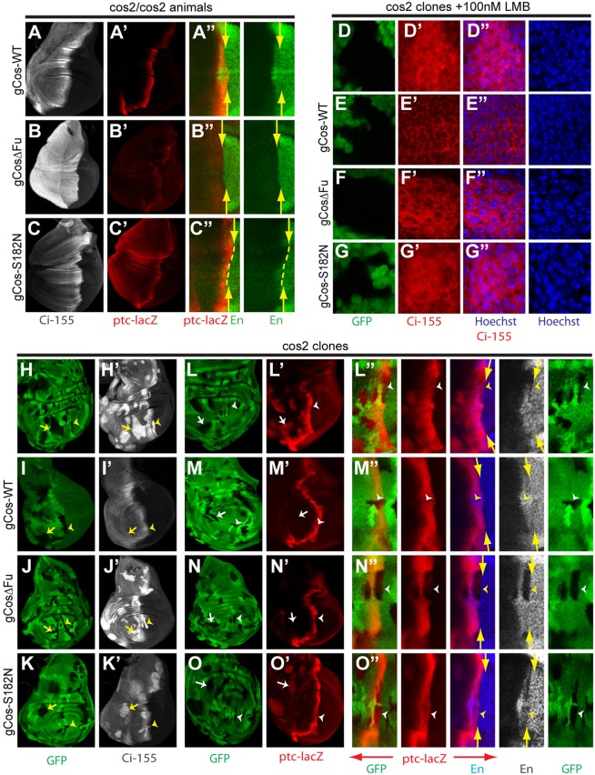
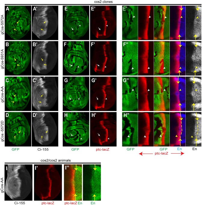
The Drosophila kinesin-family protein Costal 2 (Cos2) and its mammalian ortholog Kif7 play dual roles in Hedgehog (Hh) signaling. In the absence of Hh, Cos2 and Kif7 contribute to proteolytic processing and silencing of the Hh-regulated transcription factors, Drosophila Cubitus interruptus (Ci) and mammalian Gli proteins. Cos2 and Kif7 are also necessary for full activation of full-length Ci-155 and Gli transcription factors in response to Hh proteins. Here, we use classical fused alleles and transgenic Cos2 products deficient for Fused (Fu) association to show that Cos2 must bind to Fu to support efficient Ci-155 processing. Residual Ci-155 processing in the absence of Cos2-Fu interaction did not require Suppressor of Fused, which has been implicated in processing mammalian Gli proteins. We also provide evidence that Cos2 binding to the CORD domain of Ci-155 contributes to both Ci-155 processing and Ci-155 silencing in the absence of Hh. In the presence of Hh, Ci-155 processing is blocked and Cos2 now promotes activation of Ci-155, which requires Fu kinase activity. Here, we show that normal Ci-155 activation by Hh requires Cos2 binding to Fu, supporting the hypothesis that Cos2 mediates the apposition of Fu molecules suitable for cross-phosphorylation and consequent full activation of Fu kinase. We also find that phosphorylation of Cos2 by Fu at two previously mapped sites, S572 and S931, which is thought to mediate Ci-155 activation, is not required for normal activation of Ci-155 by Hh or by activated Fu.

Keywords: Costal 2 (Cos); Drosophila; Fused; Hedgehog; Signal Transduction.


Figures

Fig. 1.
Fig. 1.
The C-terminal of Fused is required for efficient Ci-155 processing. (A) Schematic of proteins encoded by fused alleles used. (B-E) Full-length Ci-155 (red) and (B′-E′) ptc-lacZ reporter of Ci activity (green) in wing discs from male wild-type (WT) or fu mutant larvae. Arrows indicate the anterior (left) boundary of ptc-lacZ expression. Plots of Ci-155 staining intensity (above B-E) along the AP axis were generated as described in the Materials and methods for the boxed regions. Background posterior Ci-155 levels (red line) and anterior Ci-155 levels (blue line) in wild-type discs are indicated. (F-I) Myc epitope staining (red) of Ci transgenes tagged with Myc at the C terminus and expressed ubiquitously using the C765-GAL4 driver in wild-type (WT) or fuM1 mutant wing discs. Brackets indicate the estimated AP border territory from parallel ptc-lacZ staining (not shown). (J-K′) Ci-155 levels (red) were increased in anterior fuM1 clones (white arrows) marked by two copies of a ubi-GFP transgene (adjacent to GFP-negative twin-spot clones) in otherwise normal discs (J,J′) and in discs expressing UAS-cos2 at a high level with the C765-GAL4 driver (K,K′). Excess Cos2 inhibited Hh signaling and reduced Ci-155 staining at the AP border (yellow arrows). Insets show boxed regions at higher magnification. (L-M″) hh-lacZ (red) was repressed by Ci-75 repressor derived from UAS-Ci expressed using C765-GAL4 in posterior fuM1 smo clones (arrows), marked by loss of Fu staining (green) in otherwise normal discs (L-L″) and in homozygous Su(fu)LP (null) discs (M-M″).
Fig. 2.
Fig. 2.
Ci-155 processing by overexpressed Cos2 deficient for Fu association. (A) Kc cells were transfected with DNAs encoding Flag-tagged Cos2 proteins and HA-tagged Fu as shown, followed by immunoprecipitation with Flag antibody (IP: Flag) and western blot with Flag and HA antibodies to measure Fu association with Cos2. Numbers indicate the relative intensity of HA-Fu signals in immunoprecipitates and cell extracts. (B-C″) Fu protein levels (red) were (B-B″) greatly increased in cos2 mutant clones that express UAS-cos2 (arrows), marked by GFP (green), but (C-C″) severely reduced in cos2 mutant clones that express UAS-CosΔFu. (D-F) Cos2 levels (blue) were greatly increased in cos2 clones (arrows), marked by GFP (green), that express (E′) UAS-Cos2 or (F′) UAS-CosΔFu compared with (D′) cos2 clones alone. Increased Ci-155 (red) in (D″) cos2 mutant clones was prevented by expression of (E″) UAS-Cos2 or (F″) UAS-CosΔFu. (G-I′) Ci-155 (white) was not elevated in cos2 mutant clones (arrows), marked by GFP (G-H′) or by the absence of GFP (I,I′), when UAS-CosΔFu was expressed in the clone in (G,G′) otherwise normal discs and in (H,H′) Su(fu)LP mutant discs, or (I,I′) when UAS-CosΔFu was expressed throughout Su(fu)LP mutant discs. Insets (right) show clone regions at higher magnification.
Fig. 3.
Fig. 3.
Properties of Cos2 variants deficient for Fu or Ci-CORD binding when expressed at physiological levels. (A-C″) Wing discs homozygous for cos22 with one copy of genomic transgenes for (A) wild-type Cos2, (B) CosΔFu or (C) Cos2-S182N, showing (A-C) Ci-155 staining (white), (A′-C′) ptc-lacZ staining (red) and (A″-C″) En staining (green) alone (right) or together with ptc-lacZ (red, left) to reveal the exact position of the AP border (arrows and dashed lines) as the posterior (right) edge of ptc-lacZ staining. (D-G″) Wing discs with cos2 mutant clones marked by loss of GFP (green, D-G) and carrying the indicated transgenes, treated with 100 nM LMB for 2 h. Ci-155 (red, D′-G′) is largely absent from nuclei (blue Hoechst staining, D″-G″) in clones expressing Cos-WT (E′) or CosΔFu (F′) but not Cos2-S182N (G′) or no gCos2 transgene (D′). (H-O″) Wing discs with cos2 mutant clones, marked by loss of GFP (green) and (H,L) no Cos2 transgene or one copy of a genomic transgene for (I,M) wild-type Cos2, (J,N) CosΔFu or (K,O) Cos2-S182N, showing (H′-K′) Ci-155 staining (white) and (L′-O′) ptc-lacZ staining (red). (L″-O″) Higher magnifications of the AP border in the wing pouch are shown for (from left to right) ptc-lacZ (red) and GFP, ptc-lacZ alone, ptc-lacZ and En (blue), En alone (white), and GFP (green) alone. The AP border is marked with arrows (from ptc-lacZ staining) and GFP-negative clones are marked by arrowheads.
Fig. 4.
Fig. 4.
Cos2 with impaired Fu binding fails to activate Fu in response to activated Smo. (A-F″) Ectopic ptc-lacZ (red) was induced in anterior GFP-positive (green) smo cos2 clones by (A-C″) UAS-SmoD1-3 or (D-F″) UAS-GAP-Fu driven by C765-GAL4 in discs carrying genomic transgenes for (A-A″,D-D″) wild-type Cos2, (B-B″,E-E″) CosΔFu or (C-C″,F-F″) Cos2-S182N. Induction of ptc-lacZ by SmoD1-3 was much weaker for (B-B″) CosΔFu than for (A-A″) Cos2-WT or (C-C″) Cos2-S182N, whereas (D-F″) ptc-lacZ induction by GAP-Fu was similar for all three Cos2 transgenes. (G) Measurement of ptc-lacZ staining in response to SmoD1-3 and GAP-Fu in the presence of the indicated gCos2 transgenes. Intensity of ptc-lacZ staining relative to the AP border calculated from five anterior clones of each genotype is displayed together with the s.e.m. (see Materials and methods).
Fig. 5.
Fig. 5.
Cos2 phosphorylation site variants support normal Hh signaling. (A-H) Wing discs with cos2 mutant clones, marked by loss of GFP (green) and one copy of a genomic transgene for (A,E) Cos2-S572A, (B,F) Cos2-S931A, (C,G) Cos2-S572AS931A (Cos2-AA) or (D,H) Cos2-S572D, showing no changes in (A′-D′) Ci-155 (white) or (E′-H′) ptc-lacZ (red) staining in anterior (arrows) or AP border clones (arrowheads). (E″-H″) Higher magnifications of the AP border in the wing pouch are shown for (from left to right) GFP (green), ptc-lacZ (red), ptc-lacZ with GFP, ptc-lacZ with En (blue) and En alone (white). The AP border is marked with arrows and dashed lines (from ptc-lacZ staining) and GFP-negative clones are marked by arrowheads. (I-I″) Wing disc homozygous for cos22 with one copy of the gCos2-AA transgene, showing (I) Ci-155 staining (white), (I′) ptc-lacZ staining (red) and (I″) En staining (green) alone (right) or (red) together with ptc-lacZ (left) to reveal the exact position of the AP border (arrows).
Fig. 6.
Fig. 6.
Cos2 phosphorylation sites are not required to respond to Fu. (A-H′) Anterior smo cos2 clones (arrows) expressing UAS-GAP-Fu, marked by GFP (green), in discs carrying genomic transgenes for (A,E) wild-type Cos2, (B,F) Cos2-S572A, (C,G) Cos2-S931A or (D,H) Cos2-AA (S572A S931A) induced (A′-D′) ectopic ptc-lacZ (red) to a similar degree. (E′-G′) Ci-155 staining (white) was mildly elevated in most clones of discs with (E′) wild-type gCos2 and (G′) gCos2-S931A, but not with (F′) gCos-S572A or (H′) gCos-AA. Insets (right) show clone regions at higher magnification.
Fig. 7.
Fig. 7.
Cos2 phosphorylation sites are not required to respond to Hh even in the absence of normal Su(fu) phosphorylation. (A,B) Su(fu) mutant wing discs with UAS-Su(fu)-5A (which lacks known Fu phosphorylation sites) expressed ubiquitously using C765-GAL4, and one copy of a genomic transgene for (A) wild-type Cos2 or (B) Cos2-AA. In cos2 mutant clones, marked by loss of GFP (green) (A′,B′), ptc-lacZ staining (red) was unchanged. (A″,B″) Higher magnifications of the AP border in the wing pouch are shown for (from left to right) GFP (green), ptc-lacZ (red), ptc-lacZ with GFP, ptc-lacZ with En (blue) and En alone (white). The AP border is marked with arrows (from ptc-lacZ staining) and GFP-negative clones are marked by arrowheads. (C,D) Wings from cos22/cos22; Su(fu)LP/Su(fu)LP animals carrying two copies of the (C) gCos-WT or (D) gCos-AA transgene on the second chromosome.

Similar articles

Cited by

References

    1. Alves G., Limbourg-Bouchon B., Tricoire H., Brissard-Zahraoui J., Lamour-Isnard C. and Busson D. (1998). Modulation of Hedgehog target gene expression by the Fused serine-threonine kinase in wing imaginal discs. Mech. Dev. 78, 17-31 10.1016/S0925-4773(98)00130-0 - DOI - PubMed
    1. Amakye D., Jagani Z. and Dorsch M. (2013). Unraveling the therapeutic potential of the Hedgehog pathway in cancer. Nat. Med. 19, 1410-1422 10.1038/nm.3389 - DOI - PubMed
    1. Ascano M. Jr, Nybakken K. E., Sosinski J., Stegman M. A. and Robbins D. J. (2002). The carboxyl-terminal domain of the protein kinase fused can function as a dominant inhibitor of hedgehog signaling. Mol. Cell. Biol. 22, 1555-1566 10.1128/MCB.22.5.1555-1566.2002 - DOI - PMC - PubMed
    1. Briscoe J. and Thérond P. P. (2013). The mechanisms of Hedgehog signalling and its roles in development and disease. Nat. Rev. Mol. Cell Biol. 14, 416-429 10.1038/nrm3598 - DOI - PubMed
    1. Claret S., Sanial M. and Plessis A. (2007). Evidence for a novel feedback loop in the Hedgehog pathway involving Smoothened and Fused. Curr. Biol. 17, 1326-1333 10.1016/j.cub.2007.06.059 - DOI - PubMed

Publication types