. 2015 Jan 29;10(1):e0116280.
doi: 10.1371/journal.pone.0116280. eCollection 2015.

A rapid optical clearing protocol using 2,2'-thiodiethanol for microscopic observation of fixed mouse brain

Affiliations

A rapid optical clearing protocol using 2,2'-thiodiethanol for microscopic observation of fixed mouse brain

Yuka Aoyagi et al. PLoS One. .

Abstract

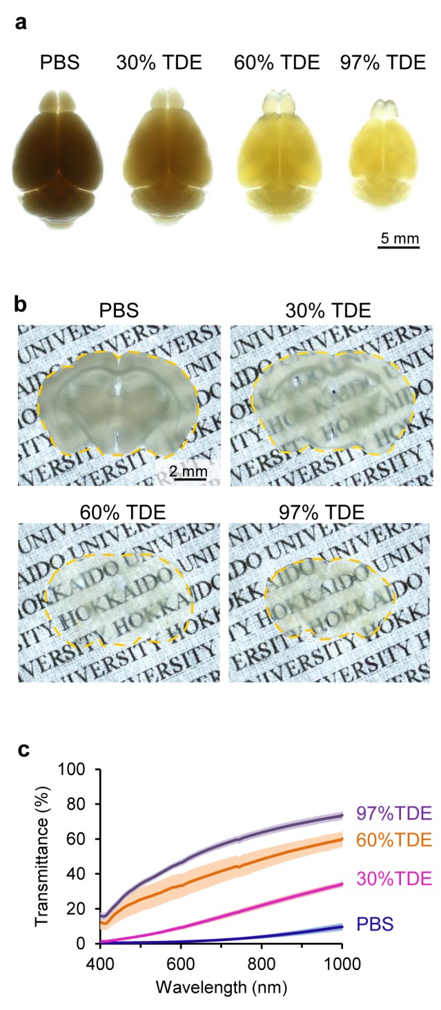
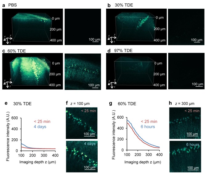
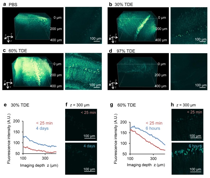
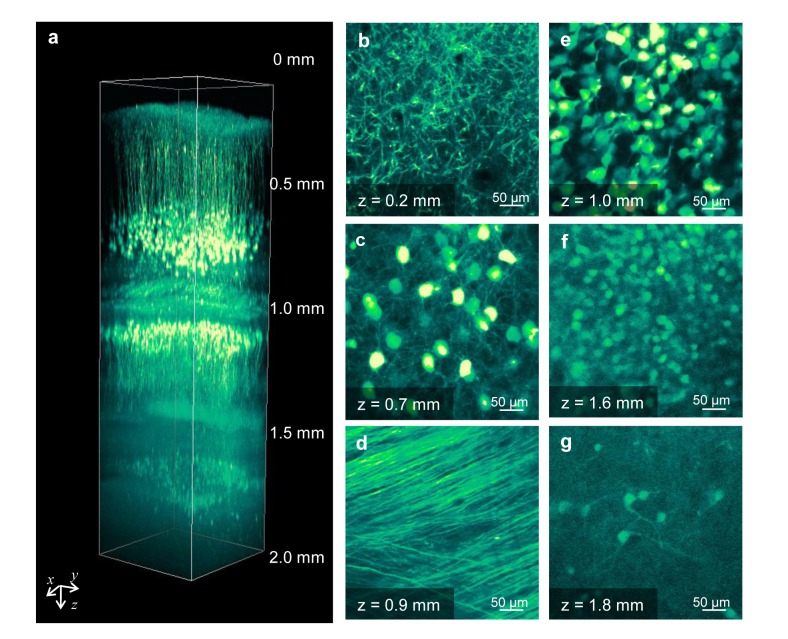
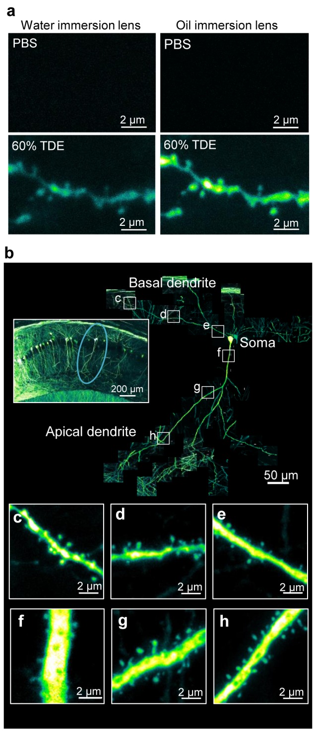
Elucidation of neural circuit functions requires visualization of the fine structure of neurons in the inner regions of thick brain specimens. However, the tissue penetration depth of laser scanning microscopy is limited by light scattering and/or absorption by the tissue. Recently, several optical clearing reagents have been proposed for visualization in fixed specimens. However, they require complicated protocols or long treatment times. Here we report the effects of 2,2'-thiodiethanol (TDE) solutions as an optical clearing reagent for fixed mouse brains expressing a yellow fluorescent protein. Immersion of fixed brains in TDE solutions rapidly (within 30 min in the case of 400-µm-thick fixed brain slices) increased their transparency and enhanced the penetration depth in both confocal and two-photon microscopy. In addition, we succeeded in visualizing dendritic spines along single dendrites at deep positions in fixed thick brain slices. These results suggest that our proposed protocol using TDE solution is a rapid and useful method for optical clearing of fixed specimens expressing fluorescent proteins.


Conflict of interest statement

Competing Interests: The authors have declared that no competing interests exist.

Figures

Figure 1
Figure 1. Fixed brain tissue clearing with TDE solutions.
(a, b) Fixed adult mouse brains after treatment with different concentrations of TDE solutions. Whole brains (a) and brain slices (400 µm in thickness) (b) were immersed in each TDE solution for 2 days and 1 h, respectively. The photograms were taken under backlighting. (c) Transmission curves of fixed brain slices (400 µm in thickness, n = 3) after 2 h of immersion in each TDE solution. Data shown represent the average ± SEM.
Figure 2
Figure 2. Enhancement of penetration depth on confocal microscopy.
(a–d) Images of YFP-expressing neurons in the hippocampal slices of thy1-YFP-H mouse in PBS (a) and after 2 h of immersion in 30% TDE (b), 60% TDE (c), and 97% TDE (d) solution. Left, three-dimensional reconstructed images; right, xy images at a depth of 200 µm from the surface. (e, f) Fluorescence intensities immediately (within 25 min) and a long time (4 days) after immersion in 30% TDE solution. (e) Plot of the mean intensity of xy images against the depth from the surface; (f) Maximum projection images within 25 min and after 4 days of immersion in 30% TDE solution. (g, h) Fluorescence intensities immediately (within 25 min) and a few hours (6 h) after immersion in 60% TDE solutions. (g) Plot of the mean intensity of xy images against the depth from the surface; (h) Maximum projection images within 25 min and after 6 h of immersion in 60% TDE solution.
Figure 3
Figure 3. Enhancement of the penetration depth on two-photon microscopy.
(a–d) Images of YFP-expressing neurons in the hippocampal slices of thy1-YFP-H mouse in PBS (a) and after 2 h of immersion in 30% TDE (b), 60% TDE (c), 97% TDE (d) solution. Left, three-dimensional reconstructed images; right, xy images at a depth of 200 µm from the slice surface. (e, f) Fluorescence intensities immediately (within 25 min) and a long time (4 days) after immersion in 30% TDE solution. (e) Plot of the mean intensity of xy images against the depth from the surface; (f) Maximum projection images within 25 min and after 4 days of immersion in 30% TDE solution. (g, h) Fluorescence intensities immediately (within 25 min) and a few hours (6 h) after immersion in 60% TDE solution. (g) Plot of the mean intensity of xy images against the depth from the surface; (h) Maximum projection images within 25 min and after 6 h of immersion in 60% TDE solution.
Figure 4
Figure 4. Two-photon deep imaging of a fixed whole brain immersed in 60% TDE.
(a) Three-dimensional reconstructed image of a whole mouse brain after 2 days of immersion in 60% TDE. (b-g) xy images at different depths from the cerebral cortex to the lower portion of the hippocampus, including apical dendrites (b) and somata (c) of layer V neurons, white matter (d), hippocampal CA1 somata (e), upper blade (f), and lower blade (g) of the hippocampal dentate gyrus (DG).
Figure 5
Figure 5. Images of dendritic spines along a single hippocampal neuron.
(a) Combination of TDE treatment and water/oil-immersion objective lens with a high NA for imaging dendritic spine shapes in deep (100 µm) regions in a fixed brain slice. (b) Connected images of dendritic spines along single pyramidal neurons extending from a depth of 30 to 100 μm from the surface of the hippocampal slice. The left inset shows a low-magnification image of the hippocampus, and the circle shows the observed neuron. (c-h) Magnified images of the dendritic spine shapes on the basal dendrite (c-e) and the apical dendrite (f-h) along the neuron shown in (b). All images are maximum projection images.

References

    1. Sporns O, Tononi G, Kötter R (2005) The human connectome: A structural description of the human brain. PLoS Comput Biol 1: e42 10.1371/journal.pcbi.0010042 - DOI - PMC - PubMed
    1. DeFelipe J (2010) From the connectome to the synaptome: an epic love story. Science 330: 1198–1201. 10.1126/science.1193378 - DOI - PubMed
    1. Kawakami R, Sawada K, Sato A, Hibi T, Kozawa Y, et al. (2013) Visualizing hippocampal neurons with in vivo two-photon microscopy using a 1030 nm picosecond pulse laser. Sci Rep 3: 1014 10.1038/srep01014 - DOI - PMC - PubMed
    1. Horton NG, Wang K, Kobat D, Clark CG, Wise FW, et al. (2013) In vivo three-photon microscopy of subcortical structures within an intact mouse brain. Nat Photonics 7: 205–209. 10.1038/nphoton.2012.336 - DOI - PMC - PubMed
    1. Kobat D, Durst ME, Nishimura N, Wong AW, Schaffer CB, et al. (2009) Deep tissue multiphoton microscopy using longer wavelength excitation. Opt Express 17: 13354–13364. 10.1364/OE.17.013354 - DOI - PubMed

Publication types

LinkOut - more resources