

Nucleus basalis of Meynert revisited: anatomy, history and differential involvement in Alzheimer's and Parkinson's disease
- PMID: 25633602
- PMCID: PMC4366544
- DOI: 10.1007/s00401-015-1392-5
Nucleus basalis of Meynert revisited: anatomy, history and differential involvement in Alzheimer's and Parkinson's disease
Abstract
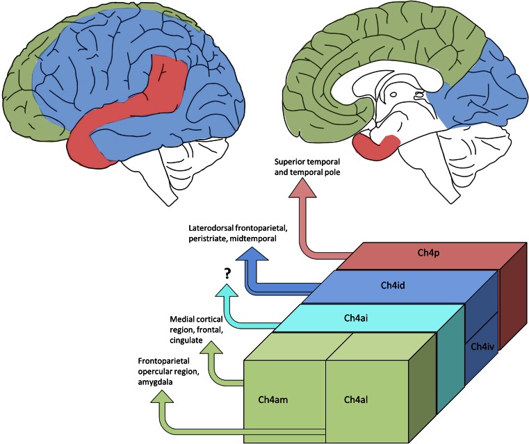
It has been well established that neuronal loss within the cholinergic nucleus basalis of Meynert (nbM) correlates with cognitive decline in dementing disorders such as Alzheimer's disease (AD). Friedrich Lewy first observed his eponymous inclusion bodies in the nbM of postmortem brain tissue from patients with Parkinson's disease (PD) and cell loss in this area can be at least as extensive as that seen in AD. There has been confusion with regard to the terminology and exact localisation of the nbM within the human basal forebrain for decades due to the diffuse and broad structure of this "nucleus". Also, while topographical projections from the nbM have been mapped out in subhuman primates, no direct clinicopathological correlations between subregional nbM and cortical pathology and specific cognitive profile decline have been performed in human tissue. Here, we review the evolution of the term nbM and the importance of standardised nbM sampling for neuropathological studies. Extensive review of the literature suggests that there is a caudorostral pattern of neuronal loss within the nbM in AD brains. However, the findings in PD are less clear due to the limited number of studies performed. Given the differing neuropsychiatric and cognitive deficits in Lewy body-associated dementias (PD dementia and dementia with Lewy bodies) as compared to AD, we hypothesise that a different pattern of neuronal loss will be found in the nbM of Lewy body disease brains. Understanding the functional significance of the subregions of the nbM could prove important in elucidating the pathogenesis of dementia in PD.
Figures


Similar articles
-
Nucleus basalis of Meynert neuronal activity in Parkinson's disease.J Neurosurg. 2019 Feb 22;132(2):574-582. doi: 10.3171/2018.11.JNS182386. Print 2020 Feb 1. J Neurosurg. 2019. PMID: 30797189
-
Delusions and visual hallucinations in a patient with Parkinson's disease with dementia showing pronounced Lewy body pathology in the nucleus basalis of Meynert.Neuropathology. 2019 Aug;39(4):319-323. doi: 10.1111/neup.12581. Epub 2019 Jun 26. Neuropathology. 2019. PMID: 31243794
-
Cholinergic deficits and galaninergic hyperinnervation of the nucleus basalis of Meynert in Alzheimer's disease and Lewy body disorders.Neuropathol Appl Neurobiol. 2020 Apr;46(3):264-278. doi: 10.1111/nan.12577. Epub 2019 Oct 1. Neuropathol Appl Neurobiol. 2020. PMID: 31454423
-
The Nucleus Basalis of Meynert and Its Role in Deep Brain Stimulation for Cognitive Disorders: A Historical Perspective.J Alzheimers Dis. 2019;69(4):905-919. doi: 10.3233/JAD-180133. J Alzheimers Dis. 2019. PMID: 31104014 Review.
-
Nucleus basalis of Meynert degeneration predicts cognitive impairment in Parkinson's disease.Handb Clin Neurol. 2021;179:189-205. doi: 10.1016/B978-0-12-819975-6.00010-8. Handb Clin Neurol. 2021. PMID: 34225962 Review.
Cited by
-
Active Inference and Auditory Hallucinations.Comput Psychiatr. 2018 Dec;2:183-204. doi: 10.1162/cpsy_a_00022. Comput Psychiatr. 2018. PMID: 30627670 Free PMC article.
-
Effects of Cigarette Smoking on Resting-State Functional Connectivity of the Nucleus Basalis of Meynert in Mild Cognitive Impairment.Front Aging Neurosci. 2021 Nov 18;13:755630. doi: 10.3389/fnagi.2021.755630. eCollection 2021. Front Aging Neurosci. 2021. PMID: 34867281 Free PMC article.
-
The Role of Leptin and Adiponectin in Obesity-Associated Cognitive Decline and Alzheimer's Disease.Front Neurosci. 2019 Jan 14;12:1027. doi: 10.3389/fnins.2018.01027. eCollection 2018. Front Neurosci. 2019. PMID: 30692905 Free PMC article. Review.
-
Effects of Prolonged Seizures on Basal Forebrain Cholinergic Neurons: Evidence and Potential Clinical Relevance.Neurotox Res. 2020 Aug;38(2):249-265. doi: 10.1007/s12640-020-00198-w. Epub 2020 Apr 21. Neurotox Res. 2020. PMID: 32319018 Review.
-
Research Mechanism and Progress of the Natural Compound Curcumin in Treating Alzheimer´s Disease.Mini Rev Med Chem. 2024;24(17):1590-1601. doi: 10.2174/0113895575263783231009051957. Mini Rev Med Chem. 2024. PMID: 37929738 Review.
References
-
- Aarsland D, Mosimann UP, McKeith IG. Role of cholinesterase inhibitors in Parkinson’s disease and dementia with Lewy bodies. J Geriatr Psychiatry Neurol. 2004;17:164–171. - PubMed
-
- Allen SJ, Dawbarn D, Wilcock GK. Morphometric immunochemical analysis of neurons in the nucleus basalis of Meynert in Alzheimer’s disease. Brain Res. 1988;454:275–281. - PubMed
-
- Angot E, Steiner JA, Hansen C, et al. Are synucleinopathies prion-like disorders? Lancet Neurol. 2010;9:1128–1138. - PubMed
-
- Arendt T, Bigl V, Arendt A, Tennstedt A. Loss of neurons in the nucleus basalis of Meynert in Alzheimer’s disease, paralysis agitans and Korsakoff’s Disease. Acta Neuropathol. 1983;61:101–108. - PubMed
-
- Arendt T, Bigl V, Tennstedt A, Arendt A. Neuronal loss in different parts of the nucleus basalis is related to neuritic plaque formation in cortical target areas in Alzheimer’s disease. Neuroscience. 1985;14:1–14. - PubMed
Publication types
MeSH terms
Grants and funding
LinkOut - more resources
Full Text Sources
Other Literature Sources
Medical
