

Reciprocal interactions between mitral valve endothelial and interstitial cells reduce endothelial-to-mesenchymal transition and myofibroblastic activation
- PMID: 25633835
- PMCID: PMC4346432
- DOI: 10.1016/j.yjmcc.2015.01.006
Reciprocal interactions between mitral valve endothelial and interstitial cells reduce endothelial-to-mesenchymal transition and myofibroblastic activation
Abstract
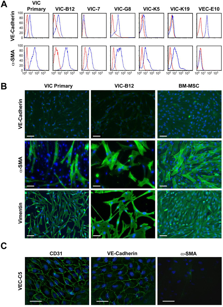
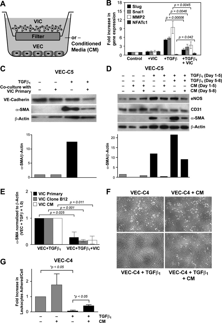
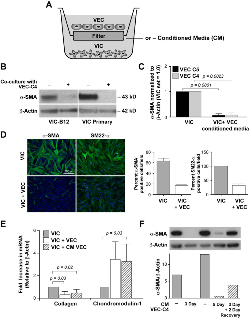
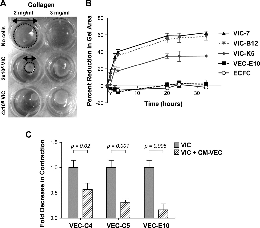
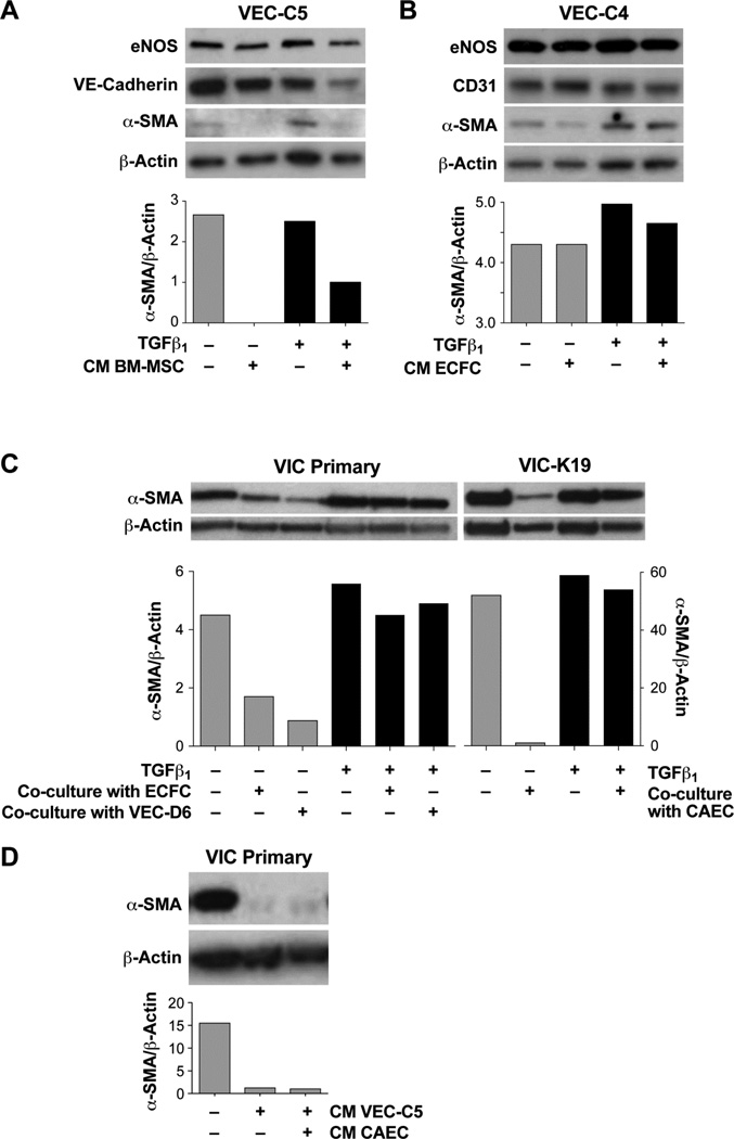
Thickening of mitral leaflets, endothelial-to-mesenchymal transition (EndMT), and activated myofibroblast-like interstitial cells have been observed in ischemic mitral valve regurgitation. We set out to determine if interactions between mitral valve endothelial cells (VECs) and interstitial cells (VICs) might affect these alterations. We used in vitro co-culture in Transwell™ inserts to test the hypothesis that VICs secrete factors that inhibit EndMT and conversely, that VECs secrete factors that mitigate the activation of VICs to a myofibroblast-like, activated phenotype. Primary cultures and clonal populations of ovine mitral VICs and VECs were used. Western blot, quantitative reverse transcriptase PCR (qPCR) and functional assays were used to assess changes in cell phenotype and behavior. VICs or conditioned media from VICs inhibited transforming growth factor β (TGFβ)-induced EndMT in VECs, as indicated by reduced expression of EndMT markers α-smooth muscle actin (α-SMA), Slug, Snai1 and MMP-2 and maintained the ability of VECs to mediate leukocyte adhesion, an important endothelial function. VECs or conditioned media from VECs reversed the spontaneous cell culture-induced change in VICs to an activated phenotype, as indicated by reduced expression of α-SMA and type I collagen, increased expression chondromodulin-1 (Chm1), and reduced contractile activity. These results demonstrate that mitral VECs and VICs secrete soluble factors that can reduce VIC activation and inhibit TGFβ-driven EndMT, respectively. These findings suggest that the endothelium of the mitral valve is critical for the maintenance of a quiescent VIC phenotype and that, in turn, VICs prevent EndMT. We speculate that the disturbance of the ongoing reciprocal interactions between VECs and VICs in vivo may contribute to the thickened and fibrotic leaflets observed in ischemic mitral regurgitation, and in other types of valve disease.
Keywords: Endothelial cells; Endothelial-to-mesenchymal transition; Mitral valve; Valve interstitial cells.
Copyright © 2015 Elsevier Ltd. All rights reserved.
Figures
Comment in
-
To 'cell' and back!J Mol Cell Cardiol. 2015 Apr;81:94-5. doi: 10.1016/j.yjmcc.2015.02.001. Epub 2015 Feb 12. J Mol Cell Cardiol. 2015. PMID: 25681583 No abstract available.
References
-
- Grande-Allen KJ, et al. Mitral valve stiffening in end-stage heart failure: evidence of an organic contribution to functional mitral regurgitation. The Journal of thoracic and cardiovascular surgery. 2005;130(3):783–790. - PubMed
-
- Grande-Allen KJ, et al. Apparently normal mitral valves in patients with heart failure demonstrate biochemical and structural derangements: an extracellular matrix and echocardiographic study. Journal of the American College of Cardiology. 2005;45(1):54–61. - PubMed
-
- Aikawa E, et al. Human semilunar cardiac valve remodeling by activated cells from fetus to adult: implications for postnatal adaptation, pathology, and tissue engineering. Circulation. 2006;113(10):1344–1352. - PubMed
Publication types
MeSH terms
Substances
Grants and funding
LinkOut - more resources
Full Text Sources
Other Literature Sources
Molecular Biology Databases
Research Materials
Miscellaneous
