. 2015 Mar;8(3):237-51.
doi: 10.1242/dmm.017830. Epub 2015 Jan 29.

Functional and molecular characterisation of EO771.LMB tumours, a new C57BL/6-mouse-derived model of spontaneously metastatic mammary cancer

Affiliations

Functional and molecular characterisation of EO771.LMB tumours, a new C57BL/6-mouse-derived model of spontaneously metastatic mammary cancer

Cameron N Johnstone et al. Dis Model Mech. 2015 Mar.

Abstract

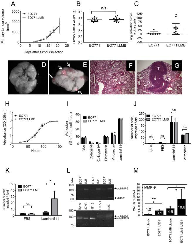
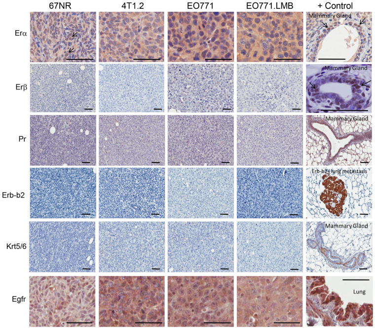
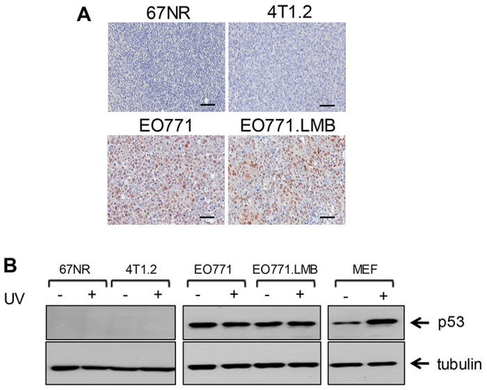
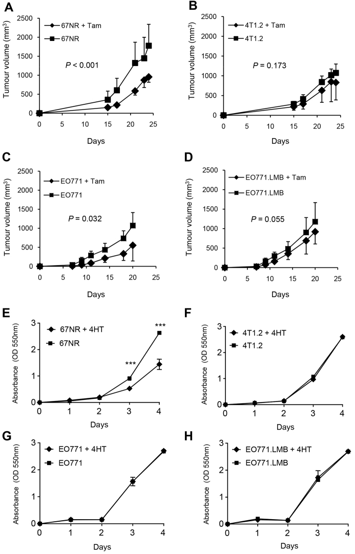
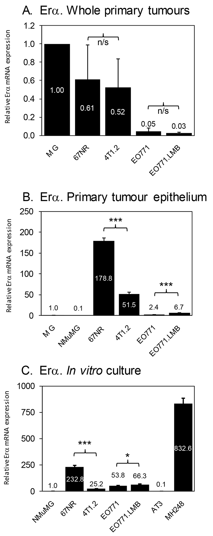
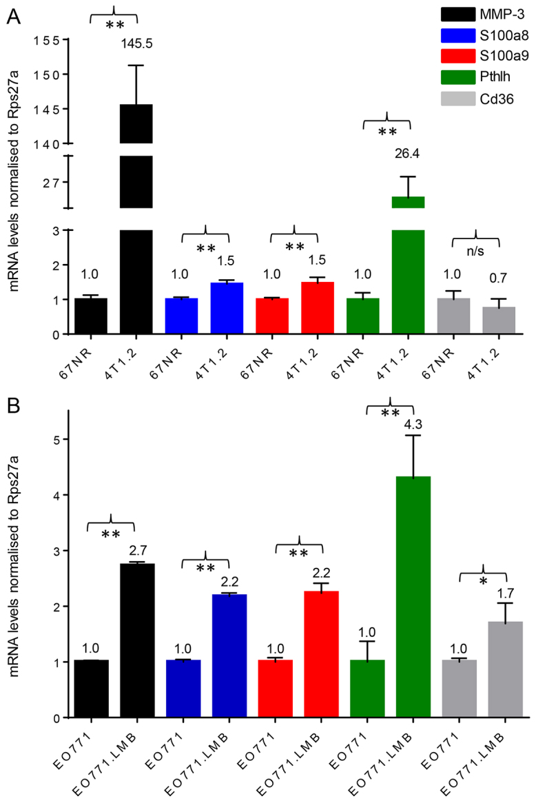
The translation of basic research into improved therapies for breast cancer patients requires relevant preclinical models that incorporate spontaneous metastasis. We have completed a functional and molecular characterisation of a new isogenic C57BL/6 mouse model of breast cancer metastasis, comparing and contrasting it with the established BALB/c 4T1 model. Metastatic EO771.LMB tumours were derived from poorly metastatic parental EO771 mammary tumours. Functional differences were evaluated using both in vitro assays and spontaneous metastasis assays in mice. Results were compared to non-metastatic 67NR and metastatic 4T1.2 tumours of the 4T1 model. Protein and transcript levels of markers of human breast cancer molecular subtypes were measured in the four tumour lines, as well as p53 (Tp53) tumour-suppressor gene status and responses to tamoxifen in vivo and in vitro. Array-based expression profiling of whole tumours identified genes and pathways that were deregulated in metastatic tumours. EO771.LMB cells metastasised spontaneously to lung in C57BL/6 mice and displayed increased invasive capacity compared with parental EO771. By immunohistochemical assessment, EO771 and EO771.LMB were basal-like, as was the 4T1.2 tumour, whereas 67NR had a luminal phenotype. Primary tumours from all lines were negative for progesterone receptor, Erb-b2/Neu and cytokeratin 5/6, but positive for epidermal growth factor receptor (EGFR). Only 67NR displayed nuclear estrogen receptor alpha (ERα) positivity. EO771 and EO771.LMB expressed mutant p53, whereas 67NR and 4T1.2 were p53-null. Integrated molecular analysis of both the EO771/EO771.LMB and 67NR/4T1.2 pairs indicated that upregulation of matrix metalloproteinase-3 (MMP-3), parathyroid hormone-like hormone (Pthlh) and S100 calcium binding protein A8 (S100a8) and downregulation of the thrombospondin receptor (Cd36) might be causally involved in metastatic dissemination of breast cancer.

Keywords: Breast cancer; Estrogen receptor alpha; Metastasis; Syngeneic preclinical models; Tumour subtyping.


Figures

Fig. 1.
Fig. 1.
Functional characterisation of the parental EO771 tumour line and its metastatic variant, EO771.LMB. (A) Parental EO771 (n=10) and metastatic variant EO771.LMB (n=5) primary tumour volumes were measured three times per week following implantation of cells into the mammary gland of C57BL/6 mice [mean tumour volume (mm3) ± s.d.]. (B) Primary tumour weights following resection on day 13 after implantation (mean weight ± s.d.; n=8, EO771; n=9, EO771.LMB). (C) Tumour burden in the lungs (mean ± s.d.) measured by qPCR quantification of mCherry DNA in genomic DNA isolated from whole lungs 2 weeks following primary tumour resection (n=8, EO771; n=9, EO771.LMB). (D,E) Superimposed brightfield/fluorescent image of lungs from EO771 (D) or EO771.LMB (E) tumour-bearing mice. Arrows indicate tumour nodules. (F,G) Hematoxylin- and eosin-stained lung sections from an EO771- (F) or EO771.LMB- (G) bearing mouse. Magnification: 40×. T: metastatic tumour deposit. (H) Proliferation of EO771 and EO771.LMB cells (mean ± s.d. of 12 replicate wells of one of three representative experiments). (I) Adhesion of EO771 and EO771.LMB cells to different substrates after 30 min. Adhesion is represented as the percentage of total cell input [mean of triplicate wells ± s.d. of a representative experiment (n=3) is shown]. (J) Chemotactic migration towards serum-free medium (SFM) or 5% (v/v) FBS after 5 hours and haptotactic migration towards laminin-511 or vitronectin after 4 hours, using Transwell inserts. The number of migrated cells was counted from three fields of view per membrane at 20× magnification (mean ± s.d. of one of three independent experiments). (K) Chemotactic and haptotactic invasion through Matrigel towards 5% (v/v) FBS or laminin-511 using Transwell inserts. The assay was run for 18 hours in triplicate wells and the number of invaded cells counted from three fields of view per membrane at 20× magnification. The data represent the mean number of invaded cells ± s.d. of one of two independent experiments. (L) Gelatin zymography of triplicate EO771 and EO771.LMB (LMB) conditioned medium from cells cultured on plastic (upper panel). The positions of molecular weight (MW) markers are shown on the left. The locations of proMMP-9 and MMP-2 are indicated on the right. Mean proMMP-9 band intensity was sixfold higher for EO771.LMB compared with EO771 (P=0.0003). Conditioned medium from primary culture of whole bone explant (bone), 67NR and 4T1.2 cells were used as positive controls for active MMP-9 (act-MMP-9) to distinguish from proMMP-9 produced by EO771 lines (lower panel). (M) qRT-PCR analysis of MMP-9 mRNA levels in mouse mammary tumour cell lines cultured on plastic or laminin-511 (LN-511, 2 μg/ml). Triplicate cultures were set up for each condition and triplicate PCR reactions run for each culture. Thus, each data point represents the mean ± s.d. from nine PCR reactions. Mean expression in EO771 on plastic was set to one. Statistical significance in B,C,I,J,K and M was determined using the Student’s t-test, whereas that in A and H were determined using two-way ANOVA. *P<0.05; **P<0.01; n/s, not significant.
Fig. 2.
Fig. 2.
Immunohistochemical staining of primary murine mammary tumours for markers of human breast cancer subtypes. From top to bottom: Luminal marker ERα. A normal mammary duct adjacent to a 67NR tumour was used as a positive control. Arrows indicate examples of ERα-positive nuclei in 67NR tumour and mammary duct. Luminal marker ERβ. A normal mammary duct was used as a positive control with arrows indicating examples of ERβ-positive nuclei. Luminal marker PR. A normal mammary gland shows high levels of this protein. Erb-b2/Neu. A metastatic tumour from the lungs of an MMTV-Neu transgenic mouse was used as a positive control. Basal markers cytokeratin 5/6 (Krt5/6) and EGFR. Positive controls (myoepithelial layer of mammary gland for Krt5/6 and mouse lung for EGFR) are shown at the right of each row. Scale bars: 50 μm.
Fig. 3.
Fig. 3.
Evaluation of p53 status. (A) Immunostaining of primary tumours for tumour suppressor p53. Scale bars: 50μm. (B) Cell lines were exposed to UVC irradiation (+) or sham-irradiated (−) and then assessed 4 hours later for p53 levels by western blot. The blots were re-probed with an antibody for α-tubulin as a loading control. UVC-treated mouse embryonic fibroblasts (MEF) with wild-type p53 were used as a positive control. The data are representative of two independent experiments.
Fig. 4.
Fig. 4.
Response of primary murine mammary tumours and cell lines to treatment with an estrogen receptor antagonist. Mice were implanted with 67NR (A), 4T1.2 (B), EO771 (C) or EO771.LMB (D) cells and divided into two groups. One group received tamoxifen (Tam, 10 μg/g of chow) beginning on the day of cell implantation and the other did not receive tamoxifen. For 67NR and 4T1.2 tumour-bearing mice, n=6/group, for EO771 and EO771.LMB, n=10/group. Graphs depict the mean tumour volume (mm3) ± s.d. for each group. (E-H) Response of cultured tumour cells to treatment with 4-hydroxytamoxifen (4HT). Cells were incubated with vehicle alone (1% ethanol) or 4HT at 500 nM. Proliferation (mean ± s.d. of six replicate wells) was measured over 4 days. One of two representative experiments is shown. ***P<0.001 by Student’s t-test.
Fig. 5.
Fig. 5.
Analysis of ERα mRNA expression. (A) qRT-PCR analysis of ERα mRNA levels in whole primary tumours. Three different primary tumours were analysed in duplicate by qRT-PCR for each tumour model. Thus, each data point represents the mean ± s.d. of six qPCR reactions across three different tumours. Expression in whole mammary gland (MG) was set to 1. (B) qRT-PCR analysis of ERα mRNA levels in whole mammary gland (MG), cultured NMuMG cells and in isolated tumour epithelial cells from the indicated primary tumours. RNA was isolated from the primary tumour cells of two mice for each tumour type and pooled. Each data point represents the mean ± s.d. of triplicate reactions. Expression in mammary gland (MG) was set to 1. (C) qRT-PCR analysis of ERα mRNA levels in cultured mammary tumour cell lines. Each data point represents the mean ± s.d. of triplicate reactions. Expression in NMuMG cells was set to 1. AT3 and MH248 murine mammary tumour lines were used as negative and positive controls, respectively. *P<0.05; ***P<0.001; n/s, not significant.
Fig. 6.
Fig. 6.
qRT-PCR analysis of MMP-3, S100a8, S100a9, Pthlh and Cd36 mRNA levels in isolated primary tumour cells. RNA was isolated from the primary tumour epithelium of two mice for each tumour type and pooled. Each data point represents the mean ± s.d. of triplicate reactions. For the pairwise comparisons, gene expression levels in non-metastatic 67NR and EO771 were set to 1 for each gene analysed. (A) 67NR and 4T1.2. (B) EO771 and EO771.LMB. *P<0.05; **P<0.01; n/s, not significant.

References

    1. Acharyya S., Oskarsson T., Vanharanta S., Malladi S., Kim J., Morris P. G., Manova-Todorova K., Leversha M., Hogg N., Seshan V. E., et al. (2012). A CXCL1 paracrine network links cancer chemoresistance and metastasis. Cell 150, 165–178. - PMC - PubMed
    1. Antoniou A. C., Kuchenbaecker K. B., Soucy P., Beesley J., Chen X., McGuffog L., Lee A., Barrowdale D., Healey S., Sinilnikova O. M., et al. CIMBA, SWE-BRCA; HEBON; EMBRACE; GEMO Collaborators Study; kConFab Investigators (2012). Common variants at 12p11, 12q24, 9p21, 9q31.2 and in ZNF365 are associated with breast cancer risk for BRCA1 and/or BRCA2 mutation carriers. Breast Cancer Res. 14, R33. - PMC - PubMed
    1. Arai K., Takano S., Teratani T., Ito Y., Yamada T., Nozawa R. (2008). S100A8 and S100A9 overexpression is associated with poor pathological parameters in invasive ductal carcinoma of the breast. Curr. Cancer Drug Targets 8, 243–252. - PubMed
    1. Aslakson C. J., Miller F. R. (1992). Selective events in the metastatic process defined by analysis of the sequential dissemination of subpopulations of a mouse mammary tumor. Cancer Res. 52, 1399–1405. - PubMed
    1. Atkinson K. E. (1989). An Introduction to Numerical Analysis. New York, NY: John Wiley and Sons.

Publication types

MeSH terms