. 2015 Apr;88(1048):20140655.
doi: 10.1259/bjr.20140655. Epub 2015 Jan 30.

Reproducibility of (18)F-FDG PET uptake measurements in head and neck squamous cell carcinoma on both PET/CT and PET/MR

Affiliations

Reproducibility of (18)F-FDG PET uptake measurements in head and neck squamous cell carcinoma on both PET/CT and PET/MR

J H Rasmussen et al. Br J Radiol. 2015 Apr.

Abstract

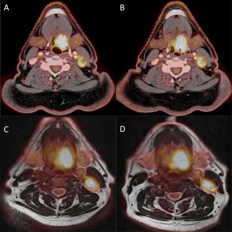
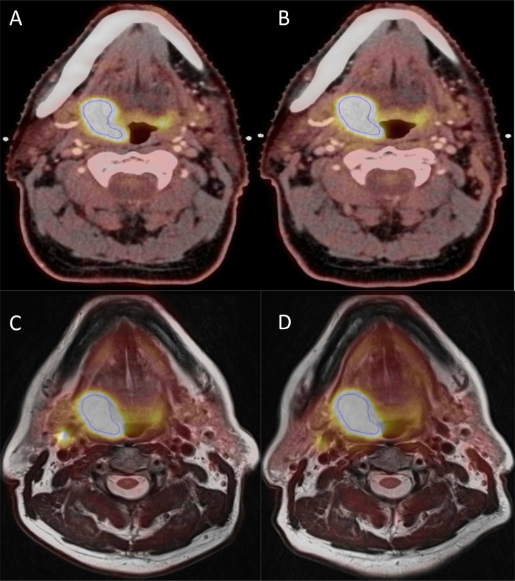
Objective: To investigate reproducibility of fluorine-18 fludeoxyglucose ((18)F-FDG) uptake on (18)F-FDG positron emission tomography (PET)/CT and (18)F-FDG PET/MR scans in patients with head and neck squamous cell carcinoma (HNSCC).

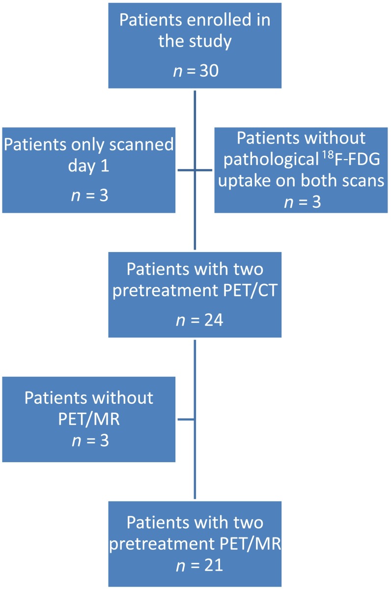
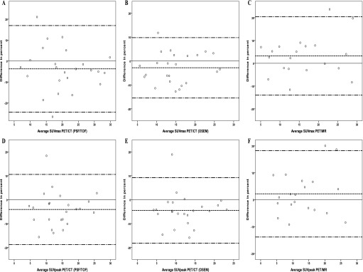
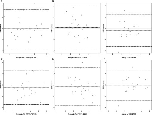
Methods: 30 patients with HNSCC were included in this prospective study. The patients were scanned twice before radiotherapy treatment with both PET/CT and PET/MR. Patients were scanned on the same scanners, 3 days apart and according to the same protocol. Metabolic tumour activity was measured by the maximum and peak standardized uptake value (SUVmax and SUVpeak, respectively), and total lesion glycolysis from the metabolic tumour volume defined from ≥50% SUVmax. Bland-Altman analysis with limits of agreement, coefficient of variation (CV) from the two modalities were performed in order to test the reproducibility. Furthermore, CVs from SUVmax and SUVpeak were compared. The area under the curve from cumulative SUV-volume histograms were measured and tested for reproducibility of the distribution of (18)F-FDG uptake.

Results: 24 patients had two pre-treatment PET/CT scans and 21 patients had two pre-treatment PET/MR scans available for further analyses. Mean difference for SUVmax, peak and mean was approximately 4% for PET/CT and 3% for PET/MR, with 95% limits of agreement less than ±20%. CV was small (5-7%) for both modalities. There was no significant difference in CVs between PET/CT and PET/MR (p = 0.31). SUVmax was not more reproducible than SUVpeak (p = 0.09).

Conclusion: (18)F-FDG uptake in PET/CT and PET/MR is highly reproducible and we found no difference in reproducibility between PET/CT and PET/MR.

Advances in knowledge: This is the first report to test reproducibility of PET/CT and PET/MR.


Figures

Figure 1.
Figure 1.
Flowchart illustrating patient inclusion and exclusion. F-FDG, fluorine-18 fludeoxyglucose; PET, positron emission tomography.
Figure 2.
Figure 2.
Bland–Altman plots of the maximum standardized uptake value (SUVmax) and peak standardized uptake value (SUVpeak) for positron emission tomography (PET)/CT reconstructed with point spread function (PSF)/time of flight (TOF) (a, d) and ordered subset expectation maximization (OSEM) (b, e) and for PET/MR (c, f). The difference in percent is plotted against the average; the solid line is reference for no difference, and the dashed lines represent the mean and the upper and lower limits of agreement
Figure 3.
Figure 3.
Bland–Altman plots of metabolic tumour volume from threshold of maximum standardize uptake value (MTV50%) and total lesion glycolysis (TLG) for positron emission tomography (PET)/CT reconstructed with point spread function (PSF)/time of flight (TOF) (a, d) and ordered subset expectation maximization (OSEM) (b, e) and for PET/MR (c, f). MTV50% and TLG were log (Ln) transformed, the y-axis is log transformed to ease the assessment of limits of agreement. The solid line is reference for no difference, and the dashed lines represent the mean and the upper and lower limits of agreement.
Figure 4.
Figure 4.
An example each from the positron emission tomography (PET)/CT (point spread function) and PET/MR scans of a patient with 35% difference in metabolic tumour volume (MTV50%) (from 9.5 to 6.1 cm3) and 27% in maximum standardized uptake value (SUVmax) (from 14.7 to 11.8) in PET/CT. In PET/MR, the difference in MTV50% was 3% (from 11.5 to 11.2 cm3) and 2% in SUVmax (from 12.1 to 12.4). (a, b) A PET/CT slice from the first and second scans, respectively, and (c, d) a PET/MR slice each from the first and second scans, respectively.
Figure 5.
Figure 5.
An example each from the positron emission tomography (PET)/CT (point spread function) and PET/MR scans of a patient with 1% difference in metabolic tumour volume (MTV50%) in PET/CT (from 9.5 to 9.6 cm3) and 11% in maximum standardized uptake value (SUVmax) (from 30.0 to 33.6). In PET/MR, the difference in MTV50% was 13% (from 12.1 to 13.7 cm3) and 4% in SUVmax (from 27.7 to 26.7). (a, b) A PET/CT slice from the first and second scans, respectively, and (c, d) a PET/MR slice each from the first and second scans, respectively.

References

    1. Xie P, Li M, Zhao H, Sun X, Fu Z, Yu J. 18F-FDG PET or PET-CT to evaluate prognosis for head and neck cancer: a meta-analysis. J Cancer Res Clin Oncol 2011; 137: 1085–93. doi: 10.1007/s00432-010-0972-y - DOI - PMC - PubMed
    1. Zhang B, Li X, Lu X. Standardized uptake value is of prognostic value for outcome in head and neck squamous cell carcinoma. Acta Otolaryngol 2010; 130: 756–62. doi: 10.3109/00016480903402981 - DOI - PubMed
    1. Rasmussen JH, Vogelius IR, Fischer BM, Friborg J, Aznar MC, Persson GF, et al. . Prognostic value of 18F-fludeoxyglucose uptake in 287 patients with head and neck squamous cell carcinoma. Head Neck May 2014. Epub ahead of print. doi: 10.1002/hed.23745 - DOI - PubMed
    1. Troost EG, Schinagl DA, Bussink J, Oyen WJ, Kaanders JH. Clinical evidence on PET-CT for radiation therapy planning in head and neck tumours. Radiother Oncol 2010; 96: 328–34. doi: 10.1016/j.radonc.2010.07.017 - DOI - PubMed
    1. Troost EG, Schinagl DA, Bussink J, Boerman OC, van der Kogel AJ, Oyen WJ, et al. . Innovations in radiotherapy planning of head and neck cancers: role of PET. J Nucl Med 2010; 51: 66–76. doi: 10.2967/jnumed.108.061499 - DOI - PubMed

Publication types

MeSH terms

Substances