. 2015 Jun:93:28-40.
doi: 10.1016/j.neuropharm.2015.01.001. Epub 2015 Jan 26.

Snake neurotoxin α-bungarotoxin is an antagonist at native GABA(A) receptors

Affiliations

Snake neurotoxin α-bungarotoxin is an antagonist at native GABA(A) receptors

Saad Hannan et al. Neuropharmacology. 2015 Jun.

Abstract

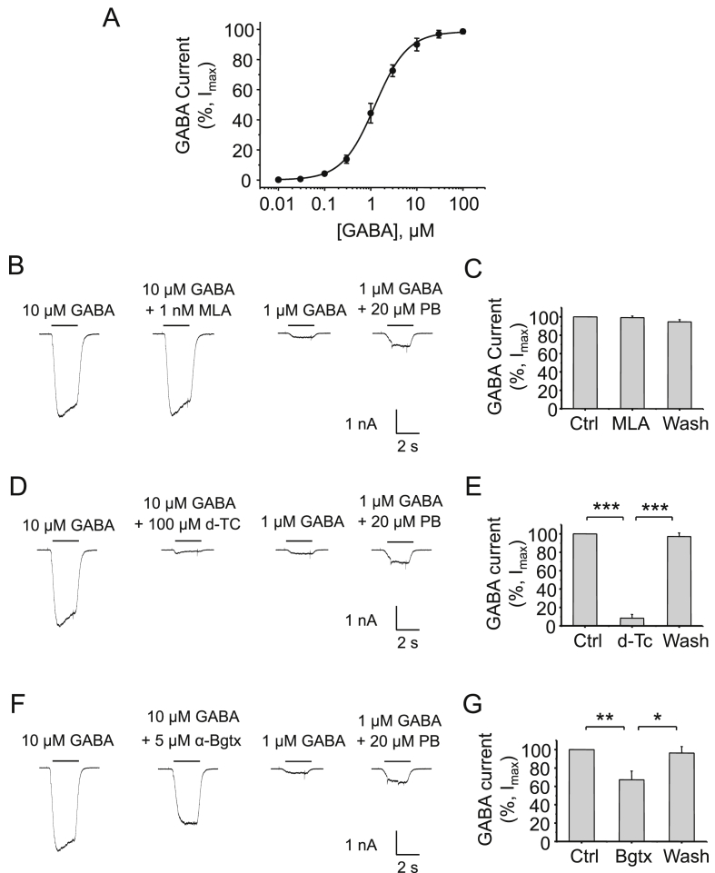
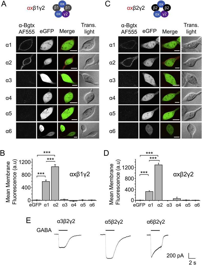
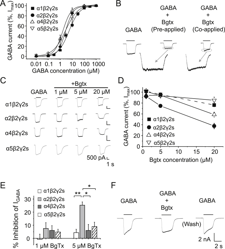
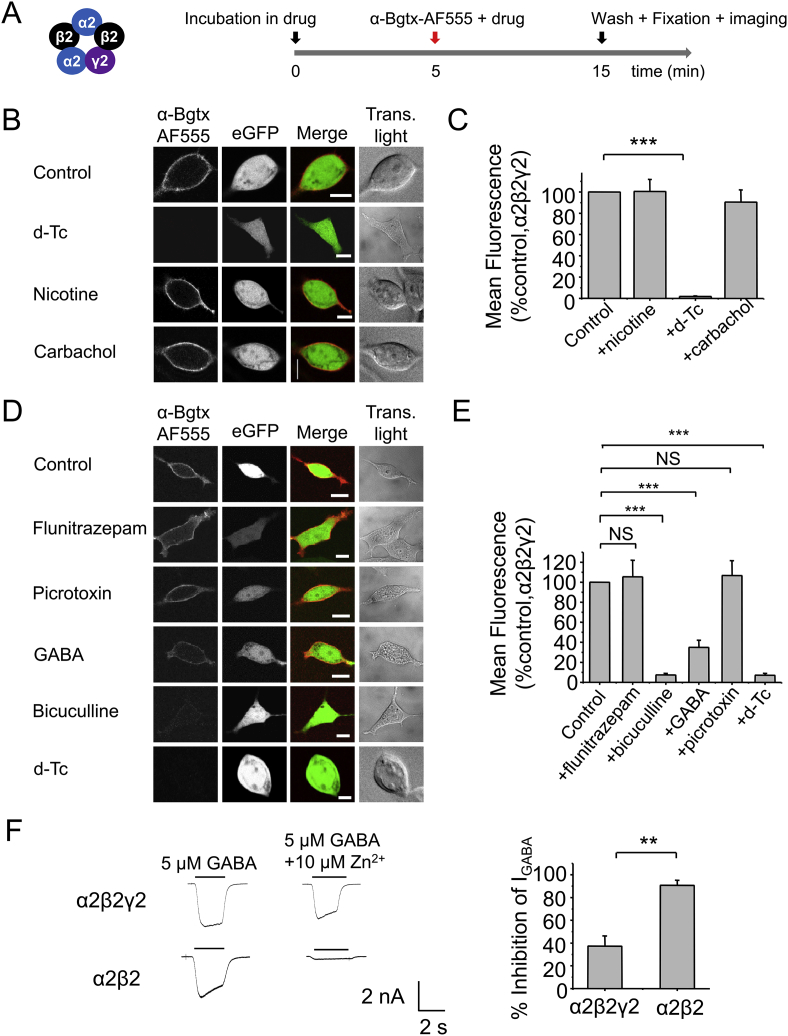
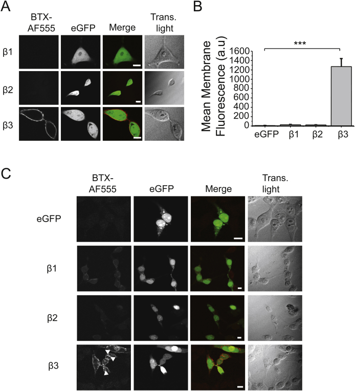
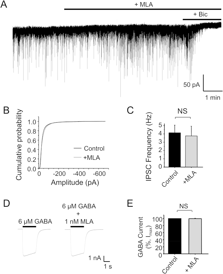
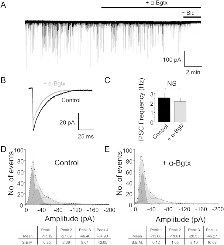
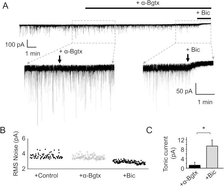
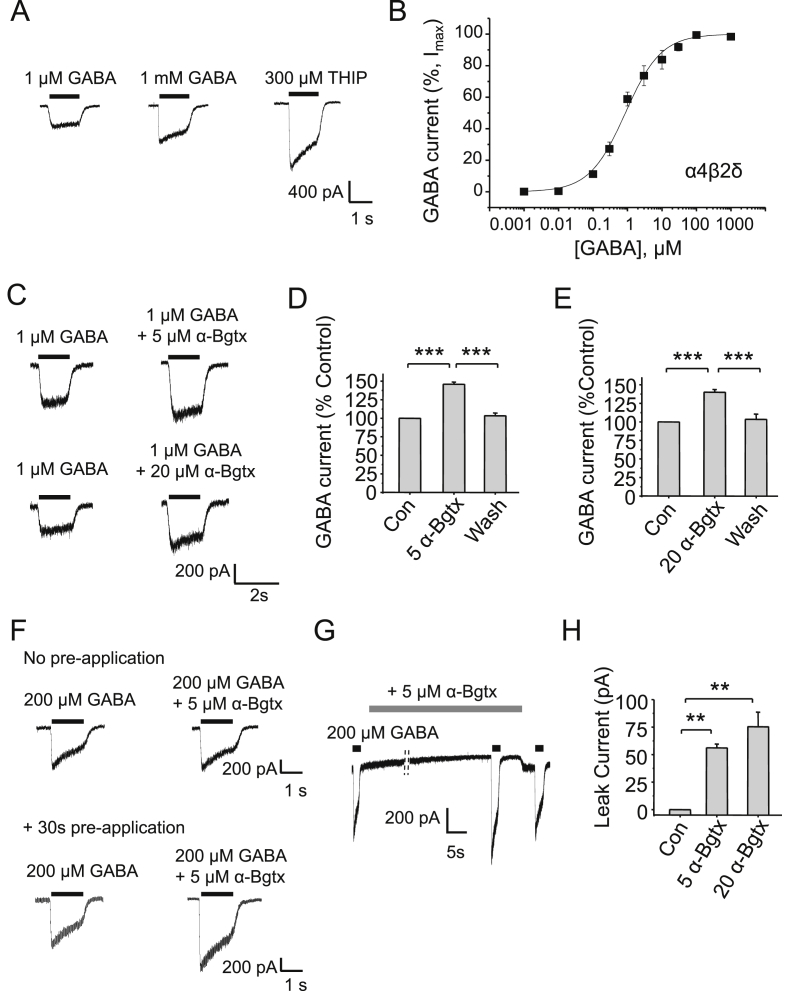
The snake neurotoxin α-bungarotoxin (α-Bgtx) is a competitive antagonist at nicotinic acetylcholine receptors (nAChRs) and is widely used to study their function and cell-surface expression. Increasingly, α-Bgtx is also used as an imaging tool for fluorophore-labelling studies, and given the structural conservation within the pentameric ligand-gated ion channel family, we assessed whether α-Bgtx could bind to recombinant and native γ-aminobutyric type-A receptors (GABAARs). Applying fluorophore-linked α-Bgtx to recombinant αxβ1/2γ2 GABAARs expressed in HEK-293 cells enabled clear cell-surface labelling of α2β1/2γ2 contrasting with the weaker staining of α1/4β1/2γ2, and no labelling for α3/5/6β1/2γ2. The labelling of α2β2γ2 was abolished by bicuculline, a competitive antagonist at GABAARs, and by d-tubocurarine (d-Tc), which acts in a similar manner at nAChRs and GABAARs. Labelling by α-Bgtx was also reduced by GABA, suggesting that the GABA binding site at the receptor β-α subunit interface forms part of the α-Bgtx binding site. Using whole-cell recording, high concentrations of α-Bgtx (20 μM) inhibited GABA-activated currents at all αxβ2γ2 receptors examined, but at lower concentrations (5 μM), α-Bgtx was selective for α2β2γ2. Using α-Bgtx, at low concentrations, permitted the selective inhibition of α2 subunit-containing GABAARs in hippocampal dentate gyrus granule cells, reducing synaptic current amplitudes without affecting the GABA-mediated tonic current. In conclusion, α-Bgtx can act as an inhibitor at recombinant and native GABAARs and may be used as a selective tool to inhibit phasic but not tonic currents in the hippocampus.

Keywords: Dentate gyrus; Electrophysiology; GABA receptor; Immunofluorescence; Nicotinic acetylcholine receptor; α-bungarotoxin.


Figures

Fig. 1
Fig. 1
Inhibition of native hippocampal GABAARs by d-Tc and α-Bgtx. A, GABA concentration response curve from primary hippocampal neurons. Whole-cell GABA-activated currents recorded from rat hippocampal neurons in culture (12–14 DIV) in response to 10 μM GABA (left hand panels) and GABA + 1 nM MLA (B,C); +100 μM d-tubocurarine (d-Tc) (D,E); +5 μM α-Bgtx and 1 nM MLA (F,G). Example control traces also show the potentiation of 1 μM GABA currents with 20 μM pentobarbital. MLA and or α-Bgtx were pre-applied for 1 min before co-application with GABA. Bargraph data presented here and in succeeding figures are means ± S.E.M, n = 5–8 cells; *P < 0.05, **P < 0.01, ***P < 0.001; One-way ANOVA.
Fig. 2
Fig. 2
GABAAR heteromers bind to Alexa Fluor 555-labelled α-Bgtx. HEK-293 cells expressing eGFP, α1-6 and γ2 with either β1 (A) or β2 (C) subunits, were incubated in 400 nM α-Bgtx coupled to Alexa Fluor 555 (α-BgTx-AF555), 48 h post-transfection, for 10 min at RT. Cells were washed to remove excess α-Bgtx-AF555, fixed and imaged. (B,D) Mean cell surface fluorescence of α-Bgtx-AF555 bound to GABAARs expressing eGFP alone, or eGFP and α1-6β1γ2 (B) or α1-6β2γ2 (D). ***P < 0.001, n = 6–9 cells; Scale bar 5 μm. Example traces of whole-cell 10 μM GABA-activated currents recorded from HEK cells expressing α3/5/6β2γ2 demonstrate the functional expression of these receptors.
Fig. 3
Fig. 3
α-Bgtx inhibition at GABAARs expressed in HEK293 cells. A, GABA concentration response curves for α1β2γ2, α2β2γ2, α4β2γ2, and α5β2γ2 receptors expressed in HEK-293 cells. B, GABA current profiles for α4β2γ2 receptors in response to 3 μM GABA, and +20 μM α-Bgtx (with or without pre-application for 1 min). Inserts show expanded current profiles. C, Representative whole-cell GABA-activated currents in response to submaximal concentrations of GABA in the absence (left-panels) or presence of 1, 5 and 20 μM α-Bgtx (pre-applied for 1 min) for cells expressing α1β2γ2, α2β2γ2, α4β2γ2 and α5β2γ2 receptors. D, Inhibition of GABA-activated currents by α-Bgtx. GABA concentrations are 6 μM (α1β2γ2), and 3 μM (α2β2γ2, α4β2γ2, and α5β2γ2). Lines are drawn (n = 3). E, Inhibition at receptors by 1 and 5 μM α-Bgtx. With 5 μM α-Bgtx, the inhibition observed at α2β2γ2 was statistically significant compared to α1β2γ2 (**P < 0.01), and α4β2γ2 and α5β2γ2 (*P < 0.05) receptors, n = 3, One-way ANOVA. F, Representative whole-cell GABA-activated currents in response to maximal concentration of GABA (1 mM) in the presence of 5 μM α-Bgtx and after recovery from inhibition.
Fig. 4
Fig. 4
α-Bgtx binds at the β–α subunit interface of GABAARs. A, Schematic of the experimental protocol. 48 hrs after transfection, HEK-293 cells expressing eGFP and α2β2γ2 were incubated in drug for 5 min at RT followed by the addition of drug +400 nM α-Bgtx-AF555 for 10 min at RT to label cell surface receptors. The cells were washed to remove the excess α-Bgtx-AF555, fixed and imaged. B, Images of cells expressing α2β2γ2, stained with α-Bgtx-AF555 in the presence of 1 mM d-Tc, 1 mM nicotine, or 1 mM carbachol. C, Mean cell surface fluorescence of α-Bgtx-AF555 bound to surface GABAARs in the presence of d-Tc, nicotine or carbachol. D, Images of cells expressing α2β2γ2, stained with α-Bgtx-AF555 in the presence of 250 μM GABA, 50 μM bicuculline, 1 mM d-Tc, 500 nM flunitrazepam or 20 μM picrotoxin. E, Mean cell surface fluorescence of α-Bgtx-AF555 bound to GABAARs in the presence of: GABA, bicuculline, d-Tc, flunitrazepam, picrotoxin. F, 5 μM GABA-activated currents and mean (±sem) inhibition caused by 5 μM Zn2+ for HEK-293 cells expressing α2β2γ2 or α2β2 receptors, 48 h post-transfection. ***P < 0.001, **P < 0.01; n = 6–7. Scale bars 5 μm.
Fig. 5
Fig. 5
α-Bgtx-AF555 binds to β3 but not β1 or β2 subunits. A, Images of HEK-293 cells expressing eGFP with either β1, β2 or β3 subunits, incubated with 400 nM α-Bgtx-AF555 for 10 min at RT, 48 h after transfection, washed to remove the excess α-Bgtx-AF555, fixed and imaged. B, Mean surface membrane fluorescence of cells expressing eGFP and β1–3 subunits. ***P < 0.001, n = 6–9. C, Images of HEK-293 cells expressing eGFP with or without either β1, β2 or β3 subunits, after fixation in 4% PFA, 48 h after transfection, and permeabilised with 0.1% w/v Triton-X100, and incubated in 400 nM α-Bgtx-AF555 for 10 min at RT, washed and imaged. Arrowheads indicate intracellular structures labelled with α-Bgtx-AF555. Scale bars 5 μm (A) and 10 μm (C).
Fig. 6
Fig. 6
MLA does not inhibit basal GABA release onto dentate gyrus granule cells. A, Representative IPSC recording from a dentate gyrus granule cell in control aCSF and after 1 nM MLA. 50 μM Bicuculline (+Bic) was applied at the end of the experiment to block all IPSCs B, Cumulative probability distributions of IPSC amplitudes in control and in 1 nM MLA. C, Frequency of IPSCs in control and in 1 nM MLA. D, Representative whole-cell GABA-activated currents from HEK-293 cells expressing α2β2γ2 receptors, 48 h after transfection, in the absence (left) or presence of 1 nM MLA (right). MLA was pre-applied for 1 min before co-application with GABA. E, Bargraph showing the lack of inhibition of GABA-currents by 1 nM MLA. n = 5–6; NS – not significant.
Fig. 7
Fig. 7
α-Bgtx inhibits phasic inhibition in hippocampal neurons. A, Spontaneous IPSCs recorded from adult mouse dentate gyrus granule cells in acute slices (P115–125) in control aCSF and in the presence of 5 μM α-Bgtx (+α-Bgtx). Bicuculline (+Bic, 50 μM) was applied to confirm the GABAergic nature of the postsynaptic currents. Note: 1 nM MLA was present throughout this experiment. B, Averaged IPSCs of 1500 events showing a reduction of amplitudes from (A) in control aCSF (black) or in the presence of α-Bgtx (gray). C, Frequency of IPSCs in control aCSF (black) or in the presence of α-Bgtx (gray). NS – not significant. D, E IPSC amplitude histograms in control aCSF (D) and in the presence of α-Bgtx (E). The parameters for each of the four Gaussians used to obtain optimal fits to the data are shown below. Data in (D) and (E) contain approximately 13000 events.
Fig. 8
Fig. 8
α-Bgtx and tonic inhibition. A, Representative IPSCs recorded from dentate gyrus granule cells from acute adult hippocampal slices (P115–125) in control aCSF followed by 5 μM α-Bgtx and 1 nM MLA (+α-Bgtx). Bicuculline (50 μM) was added after α-Bgtx to block GABAARs and assess the extent of the GABA tonic current. B, Root mean square (RMS) noise from DGGCs in control, and after α-Bgtx and then in bicuculline. RMS noise was only significantly reduced in bicuculline (n = 6, P < 0.05, unpaired two-tailed t-test). C, Net changes in tonic current after application of α-Bgtx or bicuculline.
Fig. 9
Fig. 9
α-Bgtx does not inhibit GABA-activated α4β2δ currents. A, Representative whole-cell GABA-activated currents from HEK-293 cells expressing α4β2δ receptors in response to sub-maximal (1 μM) and maximal GABA concentrations (1 mM) in comparison to 300 μM THIP. B, GABA concentration response curve for α4β2δ receptors expressed in HEK-293 cells. Experiments were performed 48 h after transfection. pEC50: 6.04 ± 0.12 (n = 5) and EC50 = 0.91 μM. C, Representative whole-cell GABA-activated currents in response to submaximal (1 μM) concentrations of GABA in the absence (left-panels) and presence of 5 and 20 μM α-Bgtx (pre-applied for 30s) in HEK-293 cells. D, Potentiation of 1 μM GABA-activated current by 5 μM α-Bgtx compared to control (Con). E, Potentiation of 1 μM GABA-activated current by 20 μM α-Bgtx compared to control (Con). F, Representative whole-cell GABA-activated currents in response to a maximal GABA concentration in the absence (left panel) and presence (right panel) of 5 μM α-BgTx with (bottom panel) or without (top panel) a 30s pre-application of α-BgTx. G, Representative GABA-activated maximal currents before, during, and after application of 5 μM α-Bgtx. Note the change in leak current during α-Bgtx). H, Changes to leak current in control (Con), +5 μM and +20 μM α-Bgtx *P < 0.05, **P < 0.01, ***P < 0.001, n = 3–7, unpaired two-tailed t-test and One-way ANOVA.

Similar articles

Cited by

References

    1. Aracri P., Consonni S., Morini R., Perrella M., Rodighiero S., Amadeo A., Becchetti A. Tonic modulation of GABA release by nicotinic acetylcholine receptors in layer V of the murine prefrontal cortex. Cereb. Cortex. 2010;20:1539–1555. - PubMed
    1. Bogdanov Y., Michels G., Armstrong-Gold C., Haydon P.G., Lindstrom J., Pangalos M., Moss S.J. Synaptic GABAA receptors are directly recruited from their extrasynaptic counterparts. EMBO J. 2006;25:4381–4389. - PMC - PubMed
    1. Brady M.L., Moon C.E., Jacob T.C. Using an alpha-bungarotoxin binding site tag to study GABAA receptor membrane localization and trafficking. J. Vis. Exp. 2014;85:C51365. - PMC - PubMed
    1. Brickley S.G., Cull-Candy S.G., Farrant M. Single-channel properties of synaptic and extrasynaptic GABAA receptors suggest differential targeting of receptor subtypes. J. Neurosci. 1999;19:2960–2973. - PMC - PubMed
    1. Brown N., Kerby J., Bonnert T.P., Whiting P.J., Wafford K.A. Pharmacological characterization of a novel cell line expressing human α4β3δ GABAA receptors. Br. J. Pharmacol. 2002;136:965–974. - PMC - PubMed

Publication types

MeSH terms

LinkOut - more resources