

Novel GCH1 variant in Dopa-responsive dystonia and Parkinson's disease
- PMID: 25634433
- PMCID: PMC4379065
- DOI: 10.1016/j.parkreldis.2015.01.004
Novel GCH1 variant in Dopa-responsive dystonia and Parkinson's disease
Abstract
Background: GTP cyclohydrolase I (GCH1) mutations are the commonest cause of Dopa-responsive dystonia (DRD). Clinical phenotypes can be broad, even within a single family.
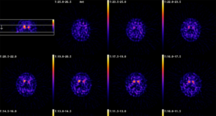
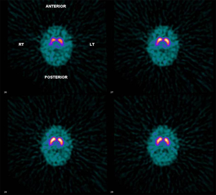
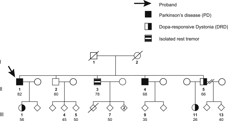
Methods: We present clinical, genetic and functional imaging data on a British kindred in which affected subjects display phenotypes ranging from DRD to Parkinson's disease (PD). Twelve family members were studied. Clinical examination, dopamine transporter (DAT) imaging, and molecular genetic analysis of GCH1 and the commonest known familial PD-related genes were performed.
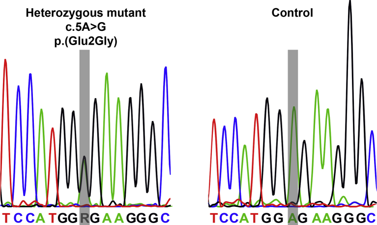
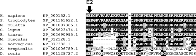
Results: We have identified a novel missense variant, c.5A > G, p.(Glu2Gly), within the GCH1 gene in affected family members displaying a range of phenotypes. Two affected subjects carrying this variant had abnormal DAT imaging. These two with abnormal DAT imaging had a PD phenotype, while the remaining three subjects with the novel GCH1 variant had normal DAT imaging and a DRD phenotype.
Conclusions: We propose that this GCH1 variant is pathogenic in this family and these findings suggest that similar mechanisms involving abnormal GTP cyclohydolase I may underlie both PD and DRD. GCH1 genetic testing should be considered in patients with PD and a family history of DRD.
Keywords: Dopa responsive dystonia; GCH1; Parkinson's disease; SPECT DAT imaging.
Copyright © 2015 The Authors. Published by Elsevier Ltd.. All rights reserved.
Figures
References
-
- Zirn B., Steinberger D., Troidi C., Brockmann K., von der Hagen M., Feiner C. Frequency of GCH1 deletions in dopa-responsive dystonia. J Neurol Neurosurg Psychiatr. 2008;79(2):183–186. - PubMed
-
- Ceravolo R., Nicoletti V., Garavaglia B., Reale C., Kiferle L., Bonuccelli U. Expanding the clinical phenotype of DYT5 mutations: is multiple system atrophy a possible one? Neurology. 2013;81(3):301–302. - PubMed
-
- Ba F., Martin W.R.W. Dopamine transporter imaging as a diagnostic tool for parkinsonism and related disorders in clinical practice. Parkinsonism Relat Disord. 2015;21(2):87–94. - PubMed
-
- Brajkovic L.D., Svetel M.V., Kostic V.S., Sobic-Saranovic D.P., Pavlovic S.V., Artiko V.M. Dopamine transporter imaging (123)I-FP-CIT (DaTSCAN) SPET in differential diagnosis of dopa-responsive dystonia and young-onset Parkinson's disease. Hell J Nucl Med. 2012;15(2):134–138. - PubMed
Publication types
MeSH terms
Substances
Supplementary concepts
Grants and funding
LinkOut - more resources
Full Text Sources
Other Literature Sources
Medical
