

DT-Diaphorase Prevents Aminochrome-Induced Alpha-Synuclein Oligomer Formation and Neurotoxicity
- PMID: 25634539
- PMCID: PMC4833033
- DOI: 10.1093/toxsci/kfv016
DT-Diaphorase Prevents Aminochrome-Induced Alpha-Synuclein Oligomer Formation and Neurotoxicity
Abstract
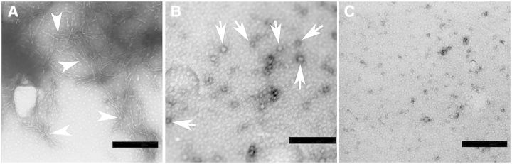
It was reported that aminochrome induces the formation of alpha synuclein (SNCA) oligomers during dopamine oxidation. We found that DT-diaphorase (NQO1) prevents the formation of SNCA oligomers in the presence of aminochrome determined by Western blot, transmission electron microscopy, circular dichroism, and thioflavin T fluorescence, suggesting a protective role of NQO1 by preventing the formation of SNCA oligomers in dopaminergic neurons. In order to test NQO1 protective role in SNCA neurotoxicity in cellular model, we overexpressed SNCA in both RCSN-3 cells (wild-type) and RCSN-3Nq7 cells, which have constitutive expression of a siRNA against NQO1. The expression of SNCA in RCSN-3SNCA and RCSN-3Nq7SNCA cells increased 4.2- and 4.4-fold, respectively. The overexpression of SNCA in RCSN-3Nq7SNCA cells induces a significant increase in cell death of 2.8- and 3.2-fold when they were incubated with 50 and 70 µM aminochrome, respectively. The cell death was found to be of apoptotic character determined by annexin/propidium iodide technique with flow cytometry and DNA laddering. A Western blot demonstrated that SNCA in RCSN-3SNCA is only found in monomer form both in the presence of 20 µM aminochrome or cell culture medium contrasting with RCSN-3Nq7SNCA cells where the majority SNCA is found as oligomer. The antioligomer compound scyllo-inositol induced a significant decrease in aminochrome-induced cell death in RCSN-3Nq7SNCA cells in comparison to cells incubated in the absence of scyllo-inositol. Our results suggest that NQO1 seems to play an important role in the prevention of aminochrome-induced SNCA oligomer formation and SNCA oligomers neurotoxicity in dopaminergic neurons.
Keywords: NQO1; Parkinsons’s disease; alpha synuclein; aminochrome; dopamine; dopaminergic neurons; neuroprotection; oligomers.
© The Author 2015. Published by Oxford University Press on behalf of the Society of Toxicology. All rights reserved. For Permissions, please e-mail: journals.permissions@oup.com.
Figures


Similar articles
-
DT-diaphorase Protects Against Autophagy Induced by Aminochrome-Dependent Alpha-Synuclein Oligomers.Neurotox Res. 2017 Oct;32(3):362-367. doi: 10.1007/s12640-017-9747-4. Epub 2017 May 6. Neurotox Res. 2017. PMID: 28478529
-
Stable expression of short interfering RNA for DT-diaphorase induces neurotoxicity.Chem Res Toxicol. 2010 Sep 20;23(9):1492-6. doi: 10.1021/tx100182a. Chem Res Toxicol. 2010. PMID: 20849151
-
Inhibition of VMAT-2 and DT-diaphorase induce cell death in a substantia nigra-derived cell line--an experimental cell model for dopamine toxicity studies.Chem Res Toxicol. 2007 May;20(5):776-83. doi: 10.1021/tx600325u. Epub 2007 Apr 11. Chem Res Toxicol. 2007. PMID: 17425337
-
Oxidation of dopamine to aminochrome as a mechanism for neurodegeneration of dopaminergic systems in Parkinson's disease. Possible neuroprotective role of DT-diaphorase.Pol J Pharmacol. 2002 Nov-Dec;54(6):573-9. Pol J Pharmacol. 2002. PMID: 12866711 Review.
-
Protective and toxic roles of dopamine in Parkinson's disease.J Neurochem. 2014 Jun;129(6):898-915. doi: 10.1111/jnc.12686. Epub 2014 Mar 18. J Neurochem. 2014. PMID: 24548101 Review.
Cited by
-
Commentary: Evaluation of Models of Parkinson's Disease.Front Neurosci. 2016 Apr 19;10:161. doi: 10.3389/fnins.2016.00161. eCollection 2016. Front Neurosci. 2016. PMID: 27147953 Free PMC article. No abstract available.
-
Aminochrome Induces Neuroinflammation and Dopaminergic Neuronal Loss: A New Preclinical Model to Find Anti-inflammatory and Neuroprotective Drugs for Parkinson's Disease.Cell Mol Neurobiol. 2023 Jan;43(1):265-281. doi: 10.1007/s10571-021-01173-5. Epub 2022 Jan 6. Cell Mol Neurobiol. 2023. PMID: 34988761 Free PMC article.
-
Amburana cearensis: Pharmacological and Neuroprotective Effects of Its Compounds.Molecules. 2020 Jul 27;25(15):3394. doi: 10.3390/molecules25153394. Molecules. 2020. PMID: 32726999 Free PMC article. Review.
-
Commentary: Gene Therapy: A Promising Approach for Neuroprotection in Parkinson's Disease?Front Neuroanat. 2017 May 19;11:40. doi: 10.3389/fnana.2017.00040. eCollection 2017. Front Neuroanat. 2017. PMID: 28579947 Free PMC article. No abstract available.
-
Neuromelanin-induced cellular stress and neurotoxicity in the pathogenesis of Parkinson's disease.Apoptosis. 2025 Aug 7. doi: 10.1007/s10495-025-02156-3. Online ahead of print. Apoptosis. 2025. PMID: 40775594 Review.
References
-
- Aguirre P., Urrutia P., Tapia V., Villa M., Paris I., Segura-Aguilar J., Núñez M. T. (2012). The dopamine metabolite aminochrome inhibits mitochondrial complex I and modifies the expression of iron transporters DMT1 and FPN1. Biometals 25, 795–803. - PubMed
-
- Arriagada C., Paris I., Sanchez de las Matas M. J., Martinez-Alvarado P., Cardenas S., Castaneda P., Graumann R., Perez-Pastene C., Olea-Azar C., Couve E., et al. (2004). On the neurotoxicity mechanism of leukoaminochrome o-semiquinone radical derived from dopamine oxidation: mitochondria damage, necrosis, and hydroxyl radical formation. Neurobiol. Dis. 16, 468–477. - PubMed
-
- Bae S. Y., Kim S., Hwang H., Kim H. K., Yoon H. C., Kim J. H., Lee S., Kim T. D. (2010). Amyloid formation and disaggregation of α-synuclein and its tandem repeat (α-TR). Biochem. Biophys. Res. Commun. 400, 531–536. - PubMed
-
- Bisaglia M., Mammi S., Bubacco L. (2007). Kinetic and structural analysis of the early oxidation products of dopamine: analysis of the interactions with alpha-synuclein. J. Biol. Chem. 282, 15597–15605. - PubMed
Publication types
MeSH terms
Substances
LinkOut - more resources
Full Text Sources
Other Literature Sources
Miscellaneous
