. 2015 Jan 27;4(1):50-66.
doi: 10.3390/biology4010050.

Treatment of leptothrix cells with ultrapure water poses a threat to their viability

Affiliations

Treatment of leptothrix cells with ultrapure water poses a threat to their viability

Tatsuki Kunoh et al. Biology (Basel). .

Abstract

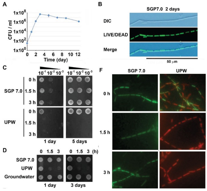
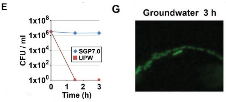
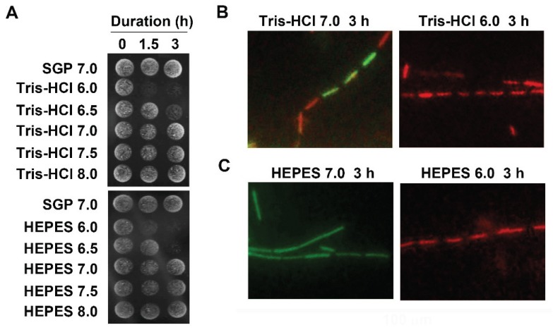
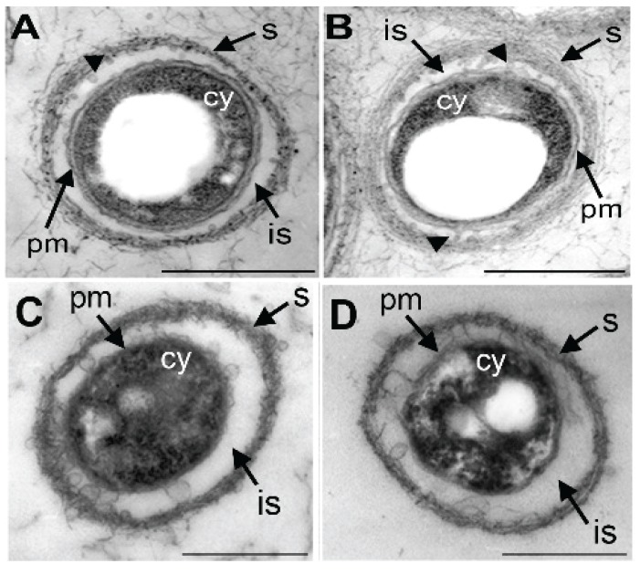
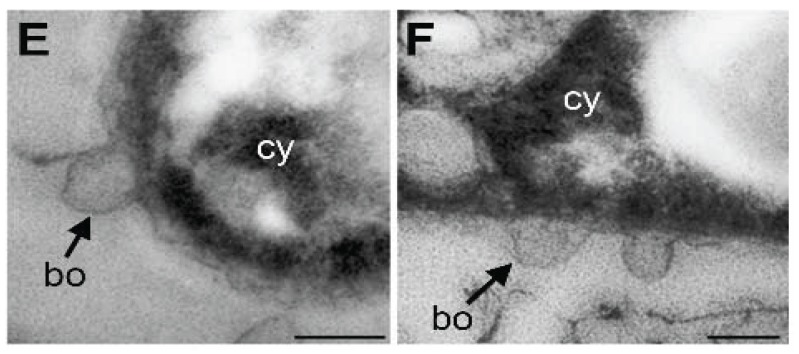
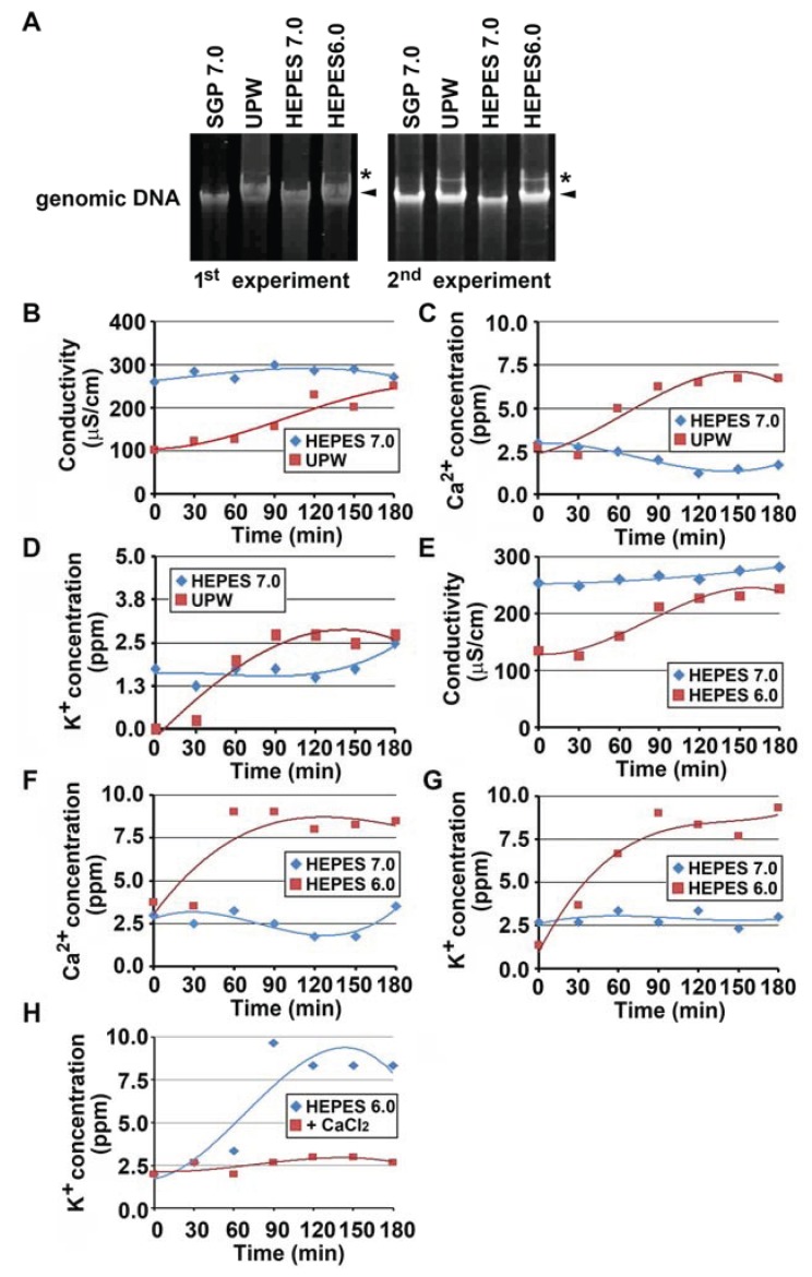
The genus Leptothrix, a type of Fe/Mn-oxidizing bacteria, is characterized by its formation of an extracellular and microtubular sheath. Although almost all sheaths harvested from natural aquatic environments are hollow, a few chained bacterial cells are occasionally seen within some sheaths of young stage. We previously reported that sheaths of Leptothrix sp. strain OUMS1 cultured in artificial media became hollow with aging due to spontaneous autolysis within the sheaths. In this study, we investigated environmental conditions that lead the OUMS1 cells to die. Treatment of the cells with ultrapure water or acidic buffers (pH 6.0) caused autolysis of the cells. Under these conditions, the plasma membrane and cytoplasm of cells were drastically damaged, resulting in leakage of intracellular electrolytes and relaxation of genomic DNA. The autolysis was suppressed by the presence of Ca2+. The hydrolysis of peptidoglycan by the lysozyme treatment similarly caused autolysis of the cells and was suppressed also by the presence of Ca2+. However, it remains unclear whether the acidic pH-dependent autolysis is attributable to damage of peptidoglycan. It was observed that L. discophora strain SP-6 cells also underwent autolysis when suspended in ultrapure water; it is however, uncertain whether this phenomenon is common among other members of the genus Leptothrix.


Figures

Figure 1
Figure 1
Autolysis of OUMS1 cells caused by UPW treatment. The cells were harvested from 2-day culture in SGP 7.0 prior to the treatment. (A) Time course of growth of OUMS1 cells in SGP7.0 determined by cfu-test. (B) Top panel: a DIC image of chained cells enveloped a thin sheath in SGP7.0. Middle panel: L/D stain image of the same chained living cells. Bottom panel: A merged image of the above DIC and L/D stain images. (C) Colony formation by OUMS1 cells treated with SGP7.0 or UPW for 0–3 h (drop test). Note no colony formation in the 1.5 or 3 h UPW treatment. (D) Colony formation by OUMS1 cells treated with SGP7.0, UPW, or GW. Note colony formation in GW and SGP7.0 treatments but no formation in UPW treatment. (E) The population of living cells in the suspension in SPG7.0 or UPW (cfu-test). Note that the living cell population reached zero in UPW by 1.5 h. (F) The L/D images of the cells incubated in SPG7.0 or UPW for 0–3 h. Note that all cells were alive in SGP7.0 treatment but some cells were dead already at 0 h UPW treatment. (G) The L/D image of the living cells treated with GW for 3 h.
Figure 1
Figure 1
Autolysis of OUMS1 cells caused by UPW treatment. The cells were harvested from 2-day culture in SGP 7.0 prior to the treatment. (A) Time course of growth of OUMS1 cells in SGP7.0 determined by cfu-test. (B) Top panel: a DIC image of chained cells enveloped a thin sheath in SGP7.0. Middle panel: L/D stain image of the same chained living cells. Bottom panel: A merged image of the above DIC and L/D stain images. (C) Colony formation by OUMS1 cells treated with SGP7.0 or UPW for 0–3 h (drop test). Note no colony formation in the 1.5 or 3 h UPW treatment. (D) Colony formation by OUMS1 cells treated with SGP7.0, UPW, or GW. Note colony formation in GW and SGP7.0 treatments but no formation in UPW treatment. (E) The population of living cells in the suspension in SPG7.0 or UPW (cfu-test). Note that the living cell population reached zero in UPW by 1.5 h. (F) The L/D images of the cells incubated in SPG7.0 or UPW for 0–3 h. Note that all cells were alive in SGP7.0 treatment but some cells were dead already at 0 h UPW treatment. (G) The L/D image of the living cells treated with GW for 3 h.
Figure 2
Figure 2
Effects of pH on induction of autolysis of OUMS1 cells. (A) Colony formation of OUMS1 cells treated with 10 mM Tris-HCl or HEPES buffers adjusted to pH 6.0–8.0 for 0–3 h, determined by drop test (3-day incubation). (B), (C) L/D stain of OUMS1 cells treated with 10 mM Tris-HCl or HEPES buffers (pH 6.0 and 7.0) for 3 h. Note that some cells died in Tris-HCl buffer even at pH 7.0 but not in HEPES7.0.
Figure 3
Figure 3
Ca2+ to suppress the acidic pH-inducing autolysis of OUMS1. (A) Colony formation of OUMS1 cells treated with SGP regardless of pH 6.0–7.5, determined by 2 day-incubation of drop-test. (B) Effects of the respective components of SGP to suppress autolysis of the cells treated with HEPES6.0, determined by 2 day-incubation of drop-test. Note that peptone and CaCl2 succeeded in suppressing the autolysis but other components did not. (C) The HEPES6.0-inducing autolysis suppressed by the presence of Ca2+ but not by Cl. (D) Suppression of HEPES6.0-inducing autolysis by higher concentrations of CaCl2. (E) HEPES6.0-inducing autolysis not suppressed by removal of Ca2+ from peptone. (F) No interference of 0.05–0.5 mM EGTA with colony formation of the cells. (G) The L/D stain showing alive and dead cells caused by 1.5 h treatment of SGP6.0, HEPES6.0, or HEPES6.0 plus CaCl2.
Figure 4
Figure 4
Figure 4. Transmission electron microscopy images of the cells treated with SGP7.0, HEPES7.0, UPW, or HEPES6.0 for 3 h. (A,B) In SGP7.0 (A) and HEPES7.0 treatments (B) the cells having double layered-plasma membrane (pm) and normal-looking cytoplasm (cy). The nearly parallel-arrayed fibrous sheath (s) formed across the intervening space (is). Globular secretion bodies seen in the intervening space in places (arrowheads). (C,D) In UPW (C) and HEPES6.0 treatments (D) the electron-dense coagulated cytoplasm (cy) surrounded hardly indistinguishable plasma membrane (pm) and electron-dense disordered sheath (s). (E,F) In UPW (E) and HEPES6.0 treatments (F) bulbous outgrowth (bo) of outer plasma membrane. Scale bar = 500 nm in (AD) and 100 nm in (E,F).
Figure 4
Figure 4
Figure 4. Transmission electron microscopy images of the cells treated with SGP7.0, HEPES7.0, UPW, or HEPES6.0 for 3 h. (A,B) In SGP7.0 (A) and HEPES7.0 treatments (B) the cells having double layered-plasma membrane (pm) and normal-looking cytoplasm (cy). The nearly parallel-arrayed fibrous sheath (s) formed across the intervening space (is). Globular secretion bodies seen in the intervening space in places (arrowheads). (C,D) In UPW (C) and HEPES6.0 treatments (D) the electron-dense coagulated cytoplasm (cy) surrounded hardly indistinguishable plasma membrane (pm) and electron-dense disordered sheath (s). (E,F) In UPW (E) and HEPES6.0 treatments (F) bulbous outgrowth (bo) of outer plasma membrane. Scale bar = 500 nm in (AD) and 100 nm in (E,F).
Figure 5
Figure 5
Relaxation of genomic DNA and electrolytes loss in OUMS1 cells treated with SGP7.0, HEPES7.0, HEPES6.0, or UPW for 3 h. (A) Genomic DNAs isolated from the cells treated with SGP7.0, UPW, HEPES7.0 or HEPES6.0 for 3 h. Arrowheads and asterisks indicate major and slowly-migrated bands, respectively. (BH) Changes of Conductivity (B,E), Ca2+ concentration (C,F), and K+ concentration (D,G,H) in the cell suspension treated with SGP7.0, UPW, HEPES7.0 or HEPES6.0 for 3 h measured every 30 min during UPW- or HEPES6.0-treatment. Plots based on triplicate means and polynomial approximation curves based on Microsoft Excel 2010 are expressed.

Similar articles

Cited by

References

    1. Suzuki T., Ishihara H., Toyoda K., Shiraishi T., Kunoh H., Takada J. Autolysis of bacterial cells leads to formation of empty sheaths by Leptothrix spp. Minerals. 2013;3:247–257. doi: 10.3390/min3020247. - DOI
    1. Spring S. The general Leptothrix and Sphaerotilus. Prokaryotes. 2006;5:758–777.
    1. Emerson D., Fleming E.J., McBeth J.M. Iron-oxidizing bacteria: An environmental and genomic perspective. Annu. Rev. Microbiol. 2010;64:561–583. doi: 10.1146/annurev.micro.112408.134208. - DOI - PubMed
    1. Ghiorse W.C. Biology of iron- and manganese-depositing bacteria. Annu. Rev. Microbiol. 1984;38:515–550. doi: 10.1146/annurev.mi.38.100184.002503. - DOI - PubMed
    1. Van Veen W.L., Mulder E.G., Deinema M.H. The Sphaerotilus-Leptothrix group of bacteria. Microbiol. Rev. 1978;42:329–356. - PMC - PubMed

LinkOut - more resources