Review
. 2015 Jan 30;116(3):515-30.
doi: 10.1161/CIRCRESAHA.116.305097.

Cellular origin and developmental program of coronary angiogenesis

Affiliations
Review

Cellular origin and developmental program of coronary angiogenesis

Xueying Tian et al. Circ Res. .

Abstract

Coronary artery disease causes acute myocardial infarction and heart failure. Identifying coronary vascular progenitors and their developmental program could inspire novel regenerative treatments for cardiac diseases. The developmental origins of the coronary vessels have been shrouded in mystery and debated for several decades. Recent identification of progenitors for coronary vessels within the endocardium, epicardium, and sinus venosus provides new insights into this question. In addition, significant progress has been achieved in elucidating the cellular and molecular programs that orchestrate coronary artery development. Establishing adequate vascular supply will be an essential component of cardiac regenerative strategies, and these findings raise exciting new strategies for therapeutic cardiac revascularization.

Keywords: coronary vessels; endocardium; sinus venosus.


Figures

Figure 1.
Figure 1.. Cre-loxP mediated genetic lineage tracing system.
A, Cre-loxP mediated recombination for lineage tracing is heritable and irreversible. In Cre expressing cells (eg. cell A), the loxP-flanked transcriptional stop cassette is removed, permitting reporter (e.g. GFP) expression in cell A and its descendants. Cre is driven by promoter a, which is strong in cell A but absent in cell B. Rosa26-loxp-stop-loxp-GFP (R26-GFP) is the Cre reporter line. B, Schematic figure showing cell A (Cre+) migrate and differentiate into cell B (Cre-). A and B cells both express lineage tracing marker GFP. C, Correct interpretation of lineage tracing data hinges on the data supporting negative Cre expression in Cell B or its non-A ancestors.
Figure 2.
Figure 2.. Formation of the nascent coronary vessel plexus in the developing heart.
A and B, The three major sources of coronary vessels, the proepicardium (PE), sinus venosus (SV) and endocardium (Endo) are intimately associated with each other during heart development. The PE is a transient structure (grey) that wedged into the atrioventricular groove between liver sinusoids and SV, and eventually gives rise to the epicardium covering the heart. The SV (blue) is the venous inflow tract. Venous cells from SV sprout onto the heart and produce subepicardial coronary vessels. The endocardium (green) lines the heart lumen. Black dashed arrows denote movement from one compartment to another, potentially complicating lineage-tracing experiments. Numbers _in B correspond to those in A showing location of migration events. C, Three putative sources for intramyocardial coronary arteries (CAs) in the developing heart. Arrows indicate corresponding migration path.
Figure 3.
Figure 3.. Epicardial contribution to developing and adult heart.
In early embryonic stage, epicardial cells form an epithelial sheet that covers the heart. At later embryonic stages, epicardial cells (Ep cells) form mesenchymal epicardium-derived cells (EPDCs) by EMT. EPDCs appear in the subepicardial layer (Sub Ep) and migrate into compact myocardium (Comp myo), where they differentiated into fibroblasts (Fb, 1), smooth muscle cells (SMC, 2), cardiomyocytes (CM, 3) and endothelial cells (EC, 4). In adult heart under normal homeostatic conditions, most epicardial cells remain quiescent (grey color). MI activates the embryonic program (red) and these epicardial cells differentiate into SMC (5) and Fb (6), but rarely if at all to CMs or ECs. Paracrine factors such as modRNA encoding Vegfa or thymosin β4 stimulates a subset of EPDCs to differentiate into EC (7) or CM (8) lineages, respectively in the post MI hearts.
Figure 4.
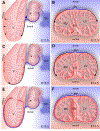
Figure 4.. Coronary vessel formation in the ventricle wall and ventricular septum.
A, C, E, Sagittal view of developing heart; B, D, F, Cross-sectional view of the developing heart. Venous cells sprout from SV and dedifferentiate into undifferentiated subepicardial ECs (black) in the dorsal side of heart. As the heart continues to develop, these subepicardial ECs penetrate the myocardial wall and differentiate into arterial ECs (red), while the remaining subepicardial ECs redifferentiate into coronary veins (blue). On the ventral side of heart and in the ventricular septum, coronary ECs arise from endocardium during trabecular compaction (black) and sprout to form a coronary plexus that connects to the coronary vessels in the ventricle wall. V, ventricle; a, atrium; SV, sinus venosus; VS, ventricular septum.
Figure 5.
Figure 5.. Contribution of SV, endocardium and epicardium to coronary vessels in the developing hearts.
Comparison of quantitative measurements from the indicated references of the contribution of different sources to coronary VECs. Upper panels show the ventricular free walls, and the lower panels show the ventricular septum. Endocardium and SV-subepicardial endothelial progenitors were the two major sources, with endocardium making the predominant contribution to VECs in the ventricular septum. The relative contribution of SV/subepicardial endothelial progenitors and endocardium to coronary vessels in the ventricular free walls is currently a matter of debate. Resolution of this uncertainty requires new genetic tools that will distinguish endcoardium from SV/subepicardidal vessels.
Figure 6.
Figure 6.. 1st and 2nd coronary vascular population in neonatal heart.
Apln-CreER, induced at E10.5, labeled fetal coronary VECs with Cre-activated RFP expression. In the P7 postnatal heart, VECs were present throughout the myocardial wall, as demonstrated by immunostaining for the VEC-selective marker FABP4 (middle panel), but the RFP lineage tracer of fetal VECs was only observed in the outer myocardial wall (left panel). The FABP4+RFP+ VECs, descended from fetal VECs, are designated the 1st coronary vascular population (CVP; red pseudocolor, right panel) while the remaining FABP4+RFP VECs, formed de novo in the postnatal heart, are designated the 2nd CVP (green pseudocolor, right panel). Bar = 1 mm.

References

    1. Wu SM, Chien KR, Mummery C. Origins and fates of cardiovascular progenitor cells. Cell 2008;132:537–543. - PMC - PubMed
    1. Hansson EM, Lindsay ME, Chien KR. Regeneration next: toward heart stem cell therapeutics. Cell Stem Cell 2009;5:364–377. - PubMed
    1. Martin-Puig S, Wang Z, Chien KR. Lives of a heart cell: tracing the origins of cardiac progenitors. Cell Stem Cell 2008;2:320–331. - PubMed
    1. Zhou B, Lin Z, Pu W. Mammalian Myocardial Regeneration. Muscle 2012;First Edition, Chapter 39:555–569.
    1. Passier R, van Laake LW, Mummery CL. Stem-cell-based therapy and lessons from the heart. Nature 2008;453:322–329. - PubMed

Publication types