Review
. 2015 Jan 22;2(1):e62.
doi: 10.1212/NXI.0000000000000062. eCollection 2015 Feb.

Does MOG Ig-positive AQP4-seronegative opticospinal inflammatory disease justify a diagnosis of NMO spectrum disorder?

Affiliations
Review

Does MOG Ig-positive AQP4-seronegative opticospinal inflammatory disease justify a diagnosis of NMO spectrum disorder?

Scott S Zamvil et al. Neurol Neuroimmunol Neuroinflamm. .

Abstract

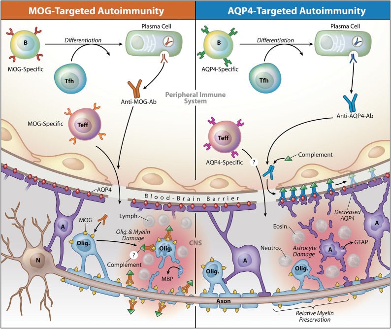
While neuromyelitis optica (NMO) immunoglobulin (Ig) G is considered the hallmark serologic marker of NMO, its association is not absolute, as NMO IgG is not detected in approximately one-fourth of the patients diagnosed with NMO spectrum disorder (NMOSD). Thus, the recent discovery that antibodies to myelin oligodendrocyte glycoprotein (MOG) are detected in some NMO IgG-seronegative patients manifesting clinical and neuroimaging signs of NMO or NMOSD has created tremendous excitement. However, it may be premature to classify this subgroup as NMOSD. NMO is considered an autoimmune astrocytopathy, and aquaporin-4 (AQP4), expressed on astrocytes, is recognized as the target autoantigen of NMO IgG. As its name denotes, MOG is produced by oligodendrocytes, CNS myelin-producing cells, and MOG is well-recognized as one of the candidate autoantigens in multiple sclerosis (MS) and acute disseminated encephalomyelitis (ADEM). Thus, is it possible that the clinical NMOSD-like phenotype associated with MOG-specific antibodies represents a variant of opticospinal MS or ADEM but not AQP4 autoimmunity or NMOSD? Whether this MOG-Ig positive AQP4-seronegative phenotype should be classified as NMOSD, opticospinal MS, or a unique entity is not simply a theoretical question but rather has practical implications for patients, their physicians, insurance carriers, and clinical investigators conducting NMO treatment trials.


Figures

Figure
Figure. Model contrasting the potential role of antibodies to myelin oligodendrocyte glycoprotein (MOG) or aquaporin-4 (AQP4) in opticospinal inflammation
MOG-specific and AQP4-specific antibodies (Ab) target 2 different CNS resident cell populations, the oligodendrocyte (Olig.) or the astrocyte (A), respectively. Data indicate that antibodies are produced outside the CNS in both MOG Ig+ AQP4-seronegative opticospinal inflammatory disease (left) and AQP4-seropositive neuromyelitis optica spectrum disease (NMOSD) (right). AQP4-specific antibodies are IgG1, an antibody subclass that requires assistance from antigen-specific T follicular helper (Tfh) cells when B cells differentiate into plasma cells. Anti-MOG antibodies appear as IgG in the diagram, although the antibody isotype is currently unknown. Serum antibodies to either MOG or AQP4 alone are not considered pathogenic in the absence of a cell-mediated inflammatory response. MOG-specific T effector cells (Teff), like in experimental autoimmune encephalitis (EAE) and possibly in multiple sclerosis (MS), or AQP4-specific Teff cells, might initiate CNS inflammation, which in AQP4-seropositive NMOSD is characterized by accumulation of neutrophils (Neutro.) and eosinophils (Eosin.). Lymphocytes (Lymph.), which are characteristic in MS and MOG-induced EAE lesions,, are shown in MOG Ig+-associated inflammation, although their presence has not been confirmed. In both conditions, inflammation may disrupt integrity of the blood-brain barrier, permitting entry of antibodies.,, MOG-specific antibodies presumably bind MOG expressed on myelin-forming oligodendrocytes and myelin, a layer that surrounds the axons extending from neuron (N) cell bodies. While the precise contribution of MOG-specific antibodies to MOG Ig+ opticospinal inflammation is yet unknown, MOG-specific antibodies promote demyelination in EAE and have been identified in MS lesions. Damage to oligodendrocytes or myelin may be associated with release of myelin basic protein (MBP). AQP4-specific IgG1 binds to AQP4 water channels, which are abundant on astrocyte end-feet processes. AQP4-specific IgG1 fixes complement, which amplifies astrocyte injury. Glial fibrillary acidic protein (GFAP) may be released upon injury to astrocytes., AQP4-targeted damage of NMOSD is associated with relative preservation of myelin. Copyright XavierStudio, reprinted with permission.

Similar articles

Cited by

References

    1. Jarius S, Wildemann B. AQP4 antibodies in neuromyelitis optica: diagnostic and pathogenetic relevance. Nat Rev Neurol 2010;6:383–392. - PubMed
    1. Pittock SJ, Lennon VA, de Seze J, et al. Neuromyelitis optica and non organ-specific autoimmunity. Arch Neurol 2008;65:78–83. - PubMed
    1. Kitley J, Waters P, Woodhall M, et al. Neuromyelitis optica spectrum disorders with aquaporin-4 and myelin-oligodendrocyte glycoprotein antibodies: a comparative study. JAMA Neurol 2014;71:276–283. - PubMed
    1. Sato DK, Callegaro D, Lana-Peixoto MA, et al. Distinction between MOG antibody-positive and AQP4 antibody-positive NMO spectrum disorders. Neurology 2014;82:474–481. - PMC - PubMed
    1. Mader S, Gredler V, Schanda K, et al. Complement activating antibodies to myelin oligodendrocyte glycoprotein in neuromyelitis optica and related disorders. J Neuroinflammation 2011;8:184. - PMC - PubMed