. 2015 Feb 5;128(3):353-60.
doi: 10.4103/0366-6999.150103.

Calreticulin translocation aggravates endoplasmic reticulum stress-associated apoptosis during cardiomyocyte hypoxia/reoxygenation

Affiliations

Calreticulin translocation aggravates endoplasmic reticulum stress-associated apoptosis during cardiomyocyte hypoxia/reoxygenation

Fei-Fei Xu et al. Chin Med J (Engl). .

Abstract

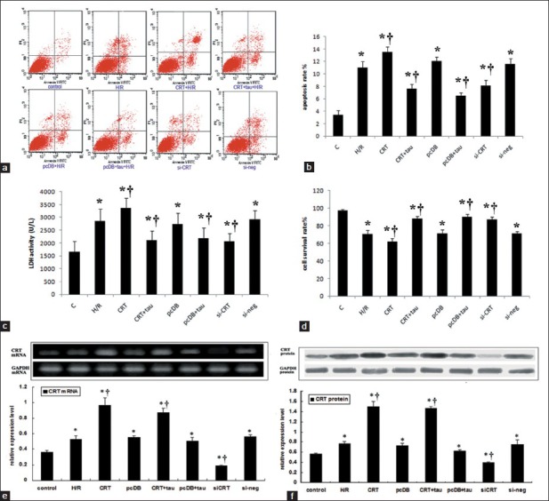
Background: Calreticulin (CRT) is major Ca 2+ -binding chaperone mainly resident in the endoplasmic reticulum (ER) lumen. Recently, it has been shown that non-ER CRT regulates a wide array of cellular responses. We previously found that CRT was up-regulated during hypoxia/reoxygenation (H/R) and this study was aimed to investigate whether CRT nuclear translocation aggravates ER stress (ERS)-associated apoptosis during H/R injury in neonatal rat cardiomyocytes.

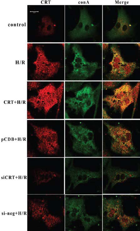
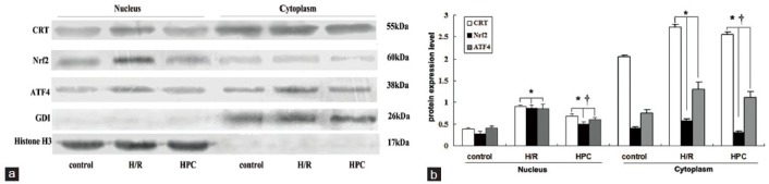
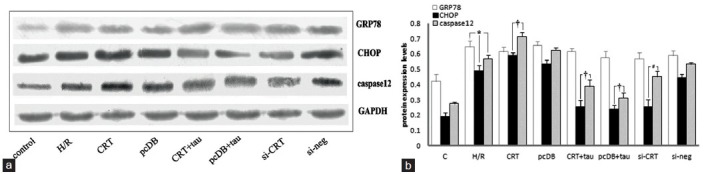
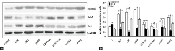
Methods: Apoptosis rate and lactate dehydrogenase (LDH) leakage in culture medium were measured as indices of cell injury. Immunofluorescence staining showed the morphological changes of ER and intracellular translocation of CRT. Western blotting or reverse transcription polymerase chain reaction was used to detect the expression of target molecules.

Results: Compared with control, H/R increased apoptosis rate and LDH activity. The ER became condensed and bubbled, and CRT translocated to the nucleus. Western blotting showed up-regulation of CRT, Nrf2, activating transcription factor 4 (ATF4), CHOP and caspase-12 expression after H/R. Exogenous CRT overexpression induced by plasmid transfection before H/R increased cell apoptosis, LDH leakage, ER disorder, CRT nuclear translocation and the expression of ERS-associated molecules. However, administration of the ERS inhibitor, taurine, or CRT siRNA alleviated cell injury, ER disorder, and inhibited ERS-associated apoptosis.

Conclusions: Our results indicated that during H/R stress, CRT translocation increases cell apoptosis and LDH leakage, aggravates ER disorder, up-regulates expression of nuclear transcription factors, Nrf2 and ATF4, and activates ERS-associated apoptosis.


Conflict of interest statement

Conflict of Interest: None declared.

Figures

Figure 1
Figure 1
The cell injuries induced by hypoxia/reoxygenation (H/R) and the effects of calreticulin (CRT) over-expression and siRNA interference and taurine administration. Cardiomyocytes were transfected by either pcDNA3.1-CRT or pcDNA3.1 or either stealth siRNA or negative control siRNA for 24 hours, then hypoxia for 8 hours and reoxygenation for 16 hours. 40 mmol/L taurine was administrated prior to hypoxia to inhibit endoplasmic reticulum stress. (a) Images of flow cytometry. (b) The statistical analysis graph of apoptosis rate analyzed by flow cytometry. (c) Lactate dehydrogenase activity of culture medium. (d) cardiomyocyte survival rate. (e) The mRNA expression and the statistical analysis graph of CRT and glyceraldehyde-3-phosphate dehydrogenase (GAPDH). (f) The protein expression and the statistical analysis graph of CRT and GAPDH (P < 0.05 vs. control, P <0.05 vs. H/R). Each experiment had been repeated for 3 times.
Figure 2
Figure 2
Co-immunocytoflurescene of concanavalin A (green) and calreticulin (CRT) (Red). During the resting state, the endoplasmic reticulum displayed a homogeneous faint green fluorescence, distributed around the nuclei. The red fluorescence representing CRT was mainly co-localized with concanavalin A, showing weak intensity in the nuclear area. After hypoxia/reoxygenation (H/R), the green fluorescence of the endoplasmic reticulum (ER) became condensed, grainy and bubbled. The fluorescence intensity of CRT increased, no longer overlapped with concanavalin A, and the low fluorescence area of the nuclei disappeared, suggesting that CRT translocated from ER to the nuclei. CRT overexpression aggravated the structural disorder of ER, and the CRT nuclear translocation became obvious. RNA interference knocked-down the total expression of CRT and the phenomena of ER bubbling and CRT nuclear translocation eventually decreased. The control plasmid (pCDB) or negative control siRNA (si-neg) transfection had no effect on ER disorder and CRT re-distribution induced by H/R. CRT + H/R represented transfection with CRT plasmid 24 hours before H/R. pcDB + H/R represented transfection with control plasmid 24 hours before H/R. siCRT + H/R represented transfection with CRT siRNA 24 hours before H/R. si-neg + H/R represented transfection with siRNA control 24 hours before H/R. The cell density for immunocytofluorescence analysis was 1 × 10⁴ cell/cm2 and the transfection efficiency of our experiment was about 65%. Each experiment was repeated in triplicate.
Figure 3
Figure 3
Cytoplasmic and nuclear extracts were separated using NE-PER nuclear and cytoplasmic extraction reagents. The relative amounts of protein expression levels of calreticulin (CRT), Nrf2, activating transcription factor 4 (ATF4). (a) Results of Western blotting of CRT, Nrf2, ATF4, Rho guanine nucleotide dissociation inhibitor-α (GDI-α) and Histone H3. Rho GDI was used as the endogenous loading control for cytoplasmic protein, and Histone H3 was used as the endogenous loading control of nuclear protein. (b) Statistical analysis of relative expression levels of nuclear and cytoplasmic proteins. The relative level of analyzed nucleus proteins was normalized to that of histone H3, and the relative level of cytoplasmic proteins was normalized to that of GDI. After hypoxia/reoxygenation (H/R), the protein expression of CRT in nuclear extracts increased compared with the control group and hypoxic preconditioning (HPC) group (P < 0.05). Hypoxic preconditioning also induced CRT nuclear translocation, but the extent was much lower than H/R group (P < 0.05) [Figure 4a and 4b]. Each experiment was repeated in triplicate (*P < 0.05 vs. control, P <0.05 vs. H/R).
Figure 4
Figure 4
The relative amounts of protein expression levels of molecular markers associated with ER stress, such as glucose-regulated protein 78 (GRP78), CHOP and caspase-12. (a) Results from Western blotting of GRP78, CHOP, caspase-12 and glyceraldehyde-3-phosphate dehydrogenase (GAPDH). (b) The statistical analysis graph of relative expression levels of these markers normalized to that of GAPDH. Hypoxia for 8 hours and reoxygenation for 16 hours (hypoxia/reoxygenation [H/R]) up-regulated the expressions of GRP78, CHOP and caspase-12. Among them, GRP78 was a protective molecule and calreticulin (CRT) over-expression, siRNA interference and taurine administration had no obvious effects on up-regulation of GRP78 induced by H/R. With regard to the pro-apoptotic molecules such as CHOP and caspase-12, CRT over-expression increased, while siRNA interference and taurine administration decreased their expression. Each experiment was repeated in triplicate. *P < 0.05 vs. control (P <0.05 vs. H/R).
Figure 5
Figure 5
The relative amounts of protein expression levels of molecular markers associated with apoptosis, such as caspase-3, Bcl-2 and Bax. (a) Results of Western blotting of caspase-3, Bcl-2, Bax and glyceraldehyde-3-phosphate dehydrogenase (GAPDH). (b) The statistical analysis graph of relative expression levels of these markers normalized to that of GAPDH. Hypoxia for 8 hours and reoxygenation for 16 hours (hypoxia/reoxygenation [H/R]) up-regulated the expressions of caspase-3 and Bax, and down-regulated Bcl-2 expression. calreticulin overexpression induced by plasmid transfection 24 h before H/R further aggravated the changes induced by H/R, while siRNA interference and taurine administration had the opposite effects. Each experiment was repeated in triplicate (*P < 0.05 vs. control. P <0.05 vs. H/R).

References

    1. Michalak M, Groenendyk J, Szabo E, Gold LI, Opas M. Calreticulin, a multi-process calcium-buffering chaperone of the endoplasmic reticulum. Biochem J. 2009;417:651–66. - PubMed
    1. Gold LI, Eggleton P, Sweetwyne MT, Van Duyn LB, Greives MR, Naylor SM, et al. Calreticulin: Non-endoplasmic reticulum functions in physiology and disease. FASEB J. 2010;24:665–83. - PMC - PubMed
    1. Mesaeli N, Nakamura K, Zvaritch E, Dickie P, Dziak E, Krause KH, et al. Calreticulin is essential for cardiac development. J Cell Biol. 1999;144:857–68. - PMC - PubMed
    1. Zwadlo C, Borlak J. Disease-associated changes in the expression of ion channels, ion receptors, ion exchangers and Ca (2+)-handling proteins in heart hypertrophy. Toxicol Appl Pharmacol. 2005;207:244–56. - PubMed
    1. Liu X, Xu F, Fu Y, Liu F, Sun S, Wu X. Calreticulin induces delayed cardioprotection through mitogen-activated protein kinases. Proteomics. 2006;6:3792–800. - PubMed

Publication types