

Positron emission tomography imaging using radiolabeled inorganic nanomaterials
- PMID: 25635467
- PMCID: PMC4540359
- DOI: 10.1021/ar500362y
Positron emission tomography imaging using radiolabeled inorganic nanomaterials
Abstract
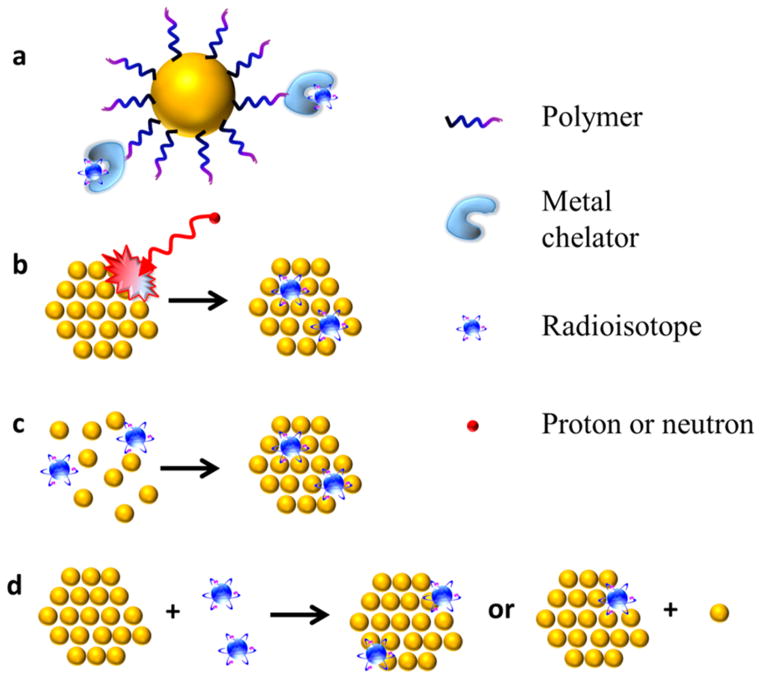
CONSPECTUS: Positron emission tomography (PET) is a radionuclide imaging technology that plays an important role in preclinical and clinical research. With administration of a small amount of radiotracer, PET imaging can provide a noninvasive, highly sensitive, and quantitative readout of its organ/tissue targeting efficiency and pharmacokinetics. Various radiotracers have been designed to target specific molecular events. Compared with antibodies, proteins, peptides, and other biologically relevant molecules, nanoparticles represent a new frontier in molecular imaging probe design, enabling the attachment of different imaging modalities, targeting ligands, and therapeutic payloads in a single vector. We introduce the radiolabeled nanoparticle platforms that we and others have developed. Due to the fundamental differences in the various nanoparticles and radioisotopes, most radiolabeling methods are designed case-by-case. We focus on some general rules about selecting appropriate isotopes for given types of nanoparticles, as well as adjusting the labeling strategies according to specific applications. We classified these radiolabeling methods into four categories: (1) complexation reaction of radiometal ions with chelators via coordination chemistry; (2) direct bombardment of nanoparticles via hadronic projectiles; (3) synthesis of nanoparticles using a mixture of radioactive and nonradioactive precursors; (4) chelator-free postsynthetic radiolabeling. Method 1 is generally applicable to different nanomaterials as long as the surface chemistry is well-designed. However, the addition of chelators brings concerns of possible changes to the physicochemical properties of nanomaterials and detachment of the radiometal. Methods 2 and 3 have improved radiochemical stability. The applications are, however, limited by the possible damage to the nanocomponent caused by the proton beams (method 2) and harsh synthetic conditions (method 3). Method 4 is still in its infancy. Although being fast and specific, only a few combinations of isotopes and nanoparticles have been explored. Since the applications of radiolabeled nanoparticles are based on the premise that the radioisotopes are stably attached to the nanomaterials, stability (colloidal and radiochemical) assessment of radiolabeled nanoparticles is also highlighted. Despite the fact that thousands of nanomaterials have been developed for clinical research, only very few have moved to humans. One major reason is the lack of understanding of the biological behavior of nanomaterials. We discuss specific examples of using PET imaging to monitor the in vivo fate of radiolabeled nanoparticles, emphasizing the importance of labeling strategies and caution in interpreting PET data. Design considerations for radiolabeled nanoplatforms for multimodal molecular imaging are also illustrated, with a focus on strategies to combine the strengths of different imaging modalities and to prolong the circulation time.
Conflict of interest statement
The authors declare no competing financial interest.
Figures
Similar articles
-
Radiolabeling Silica-Based Nanoparticles via Coordination Chemistry: Basic Principles, Strategies, and Applications.Acc Chem Res. 2018 Mar 20;51(3):778-788. doi: 10.1021/acs.accounts.7b00635. Epub 2018 Feb 28. Acc Chem Res. 2018. PMID: 29489335 Free PMC article. Review.
-
Radiolabeled inorganic nanoparticles for positron emission tomography imaging of cancer: an overview.Q J Nucl Med Mol Imaging. 2017 Jun;61(2):181-204. doi: 10.23736/S1824-4785.17.02969-7. Epub 2017 Jan 26. Q J Nucl Med Mol Imaging. 2017. PMID: 28124549 Free PMC article. Review.
-
Functionalization of inorganic nanoparticles for bioimaging applications.Acc Chem Res. 2011 Oct 18;44(10):925-35. doi: 10.1021/ar2000327. Epub 2011 Jun 7. Acc Chem Res. 2011. PMID: 21648430 Review.
-
Intrinsically radiolabeled nanoparticles: an emerging paradigm.Small. 2014 Oct 15;10(19):3825-30. doi: 10.1002/smll.201401048. Epub 2014 Jun 30. Small. 2014. PMID: 24978934 Free PMC article.
-
Chelator-Free Labeling of Metal Oxide Nanostructures with Zirconium-89 for Positron Emission Tomography Imaging.ACS Nano. 2017 Dec 26;11(12):12193-12201. doi: 10.1021/acsnano.7b05428. Epub 2017 Nov 29. ACS Nano. 2017. PMID: 29178789 Free PMC article.
Cited by
-
Diverse Applications of Nanomedicine.ACS Nano. 2017 Mar 28;11(3):2313-2381. doi: 10.1021/acsnano.6b06040. Epub 2017 Mar 14. ACS Nano. 2017. PMID: 28290206 Free PMC article. Review.
-
Understanding the in vivo Fate of Advanced Materials by Imaging.Adv Funct Mater. 2020 Sep 10;30(37):1910369. doi: 10.1002/adfm.201910369. Epub 2020 Apr 6. Adv Funct Mater. 2020. PMID: 38545084 Free PMC article.
-
Advantages and Limitations of Current Techniques for Analyzing the Biodistribution of Nanoparticles.Front Pharmacol. 2018 Aug 14;9:802. doi: 10.3389/fphar.2018.00802. eCollection 2018. Front Pharmacol. 2018. PMID: 30154715 Free PMC article. Review.
-
Recent advances in porous nanomaterials-based drug delivery systems for cancer immunotherapy.J Nanobiotechnology. 2022 Jun 14;20(1):277. doi: 10.1186/s12951-022-01489-4. J Nanobiotechnology. 2022. PMID: 35701847 Free PMC article. Review.
-
The biobehavior, biocompatibility and theranostic application of SPNS and Pd@Au nanoplates in rats and rabbits.Chem Sci. 2018 Nov 26;10(6):1677-1686. doi: 10.1039/c8sc04318c. eCollection 2019 Feb 14. Chem Sci. 2018. PMID: 30842831 Free PMC article.
References
-
- Mankoff DA. A definition of molecular imaging. J Nucl Med. 2007;48:18N–21N. - PubMed
-
- Gambhir SS. Molecular imaging of cancer with positron emission tomography. Nat Rev Cancer. 2002;2:683–693. - PubMed
-
- Ma X, Zhao Y, Liang XJ. Theranostic nanoparticles engineered for clinic and pharmaceutics. Acc Chem Res. 2011;44:1114–1122. - PubMed
-
- Cheon J, Lee JH. Synergistically integrated nanoparticles as multimodal probes for nanobiotechnology. Acc Chem Res. 2008;41:1630–1640. - PubMed
Publication types
MeSH terms
Substances
Grants and funding
LinkOut - more resources
Full Text Sources
Other Literature Sources
