. 2015 Jan;7(1):70-81.
doi: 10.18632/aging.100722.

Role of p38 mitogen-activated protein kinase in vascular endothelial aging: interaction with Arginase-II and S6K1 signaling pathway

Affiliations

Role of p38 mitogen-activated protein kinase in vascular endothelial aging: interaction with Arginase-II and S6K1 signaling pathway

Zongsong Wu et al. Aging (Albany NY). 2015 Jan.

Abstract

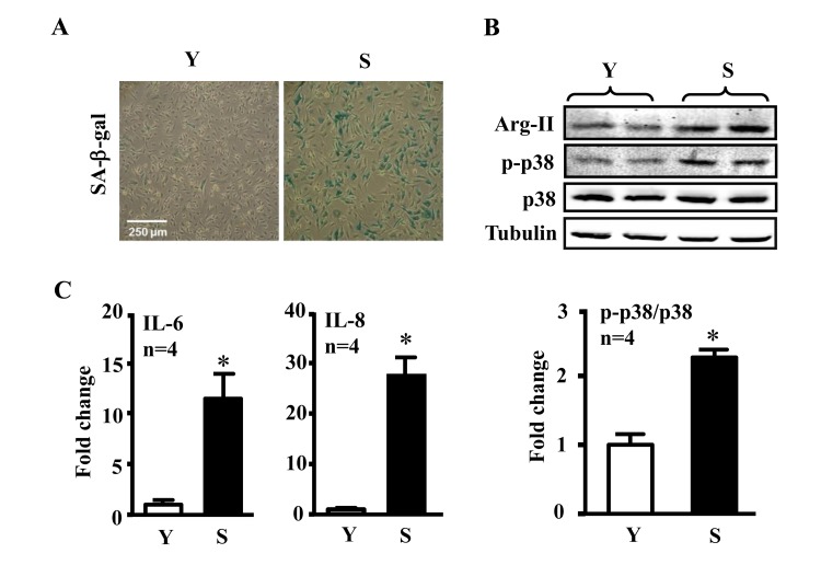
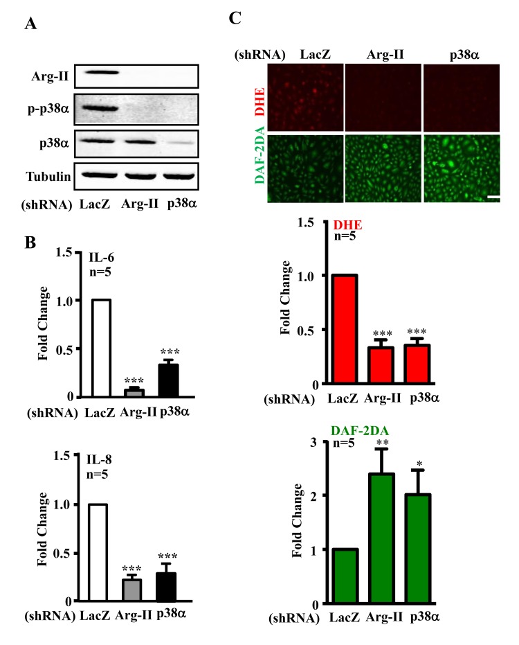
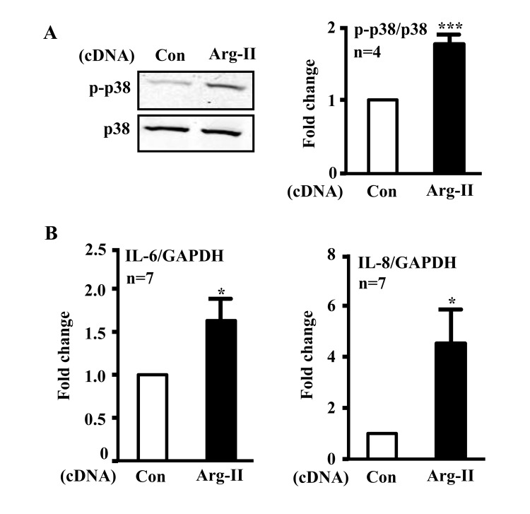
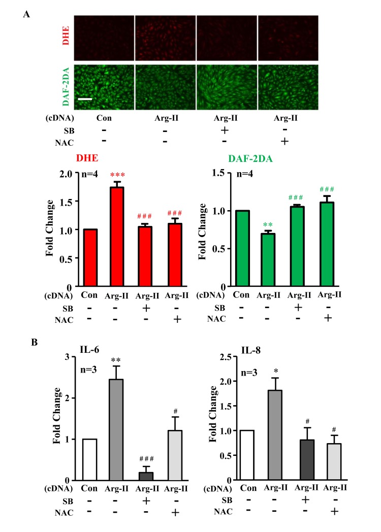
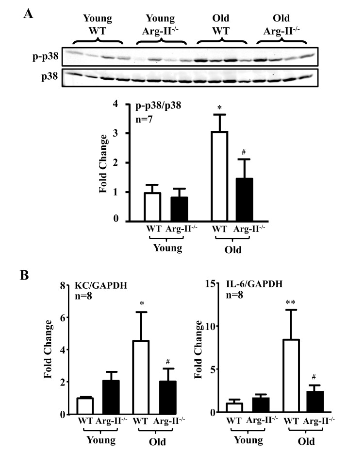
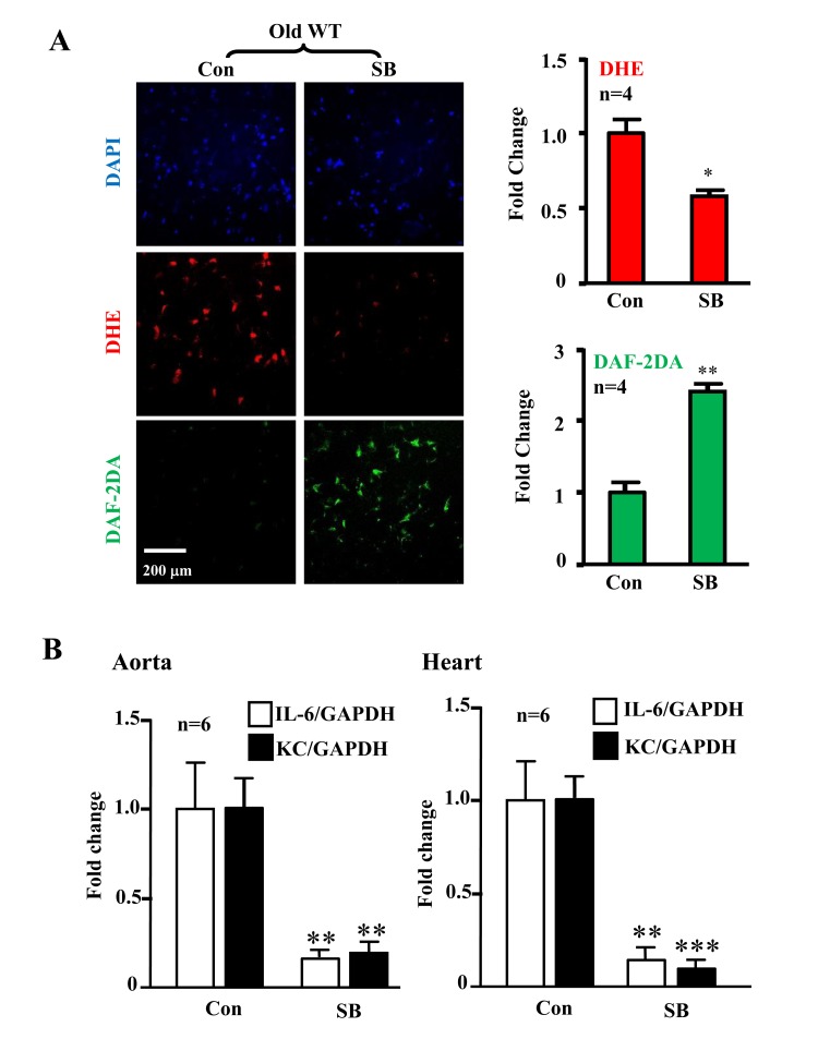
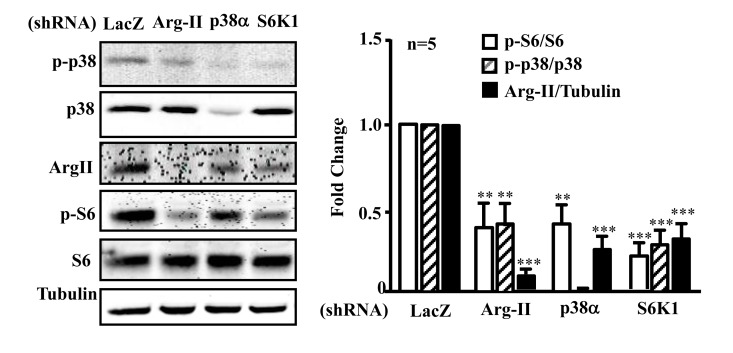
p38 mitogen-activated protein kinase (p38) regulates cellular senescence and senescence-associated secretory phenotype (SASP), i.e., secretion of cytokines and/or chemokines. Previous work showed that augmented arginase-II (Arg-II) and S6K1 interact with each other to promote endothelial senescence through uncoupling of endothelial nitric oxide synthase (eNOS). Here we demonstrate eNOS-uncoupling, augmented expression/secretion of IL-6 and IL-8, elevation of p38 activation and Arg-II levels in senescent endothelial cells. Silencing Arg-II or p38α in senescent cells recouples eNOS and inhibits IL-6 and IL-8 secretion. Overexpression of Arg-II in young endothelial cells causes eNOS-uncoupling and enhances IL-6 and IL-8 expression/secretion, which is prevented by p38 inhibition or by antioxidant. Moreover, p38 activation and expression of IL-6 and KC (the murine IL-8 homologue) are increased in the heart and/or aortas of wild type (WT) old mice, which is abolished in mice with Arg-II gene deficiency (Arg-II-/-). In addition, inhibition of p38 in the old WT mice recouples eNOS function and reduces IL-6 and KC expression in the aortas and heart. Silencing Arg-II or p38a or S6K1 inhibits each other in senescence endothelial cells. Thus, Arg-II, p38, and S6K1 form a positive circuit which regulates endothelial senescence and cardiovascular aging.


Conflict of interest statement

Conflict of interest statement

The authors declare no conflict of interest.

Figures

Figure 1
Figure 1. Comparison of inflammatory cytokines between young and senescent endothelial cells
Conditioned medium and cell lysate were collected from young (Y) and senescent (S) HUVECs which were serum-starved overnight. (A) SA-β-gal staining. (B) Immunoblotting to detect Arg-II, phsopho-p38-Thr180/Tyr182 (p-p38) and total-p38 (p38) in young and senescent cells. Tubulin served as loading control. (C) The secretion of IL-6 and IL-8 was evaluated by ELISA with collected conditioned medium described above. n=4, *p<0.05 vs young cells (Y).
Figure 2
Figure 2. Silencing Arg-II or p38α reduces cytokine/chemokine secretion and recouples eNOS in senescent endothelial cells
Senescent HUVECs were transduced with control shRNA (rAd/U6-LacZshRNA), rAd/U6-Arg-IIshRNA or -p38αshRNA. Ninety six hours post transduction, cells were serum-starved overnight. Cell culture conditioned medium were collected for ELISA, and cell extracts were subjected to immunoblotting. (A) Immunoblotting analysis of Arg-II, phsopho-p38α–Thr180/Tyr182 (p-p38α) and total-p38α(p38α). Tubulin served as loading control. (B) Cytokine/chemokine secretion was determined by ELISA with collected conditioned medium. (C) DHE and DAF-2DA staining for detection of superoxide anions and NO, respectively. Quantification of the signals were presented in the bar graphics. n=4, *p<0.05, **p<0.01, ***p<0.001 vs shRNA-LacZ group. Scale = 200 μm.
Figure 3
Figure 3. Overexpression of Arg-II in young endothelial cells activates p38 and enhances secretion of IL-6 and IL-8
HUVECs were transduced with rAd empty vector as a control or rAd/CMV-Arg-II. Forty eight hours post transduction, cells were serum-starved overnight. Cell culture conditioned medium was collected for ELISA and cell extracts were subjected to immunoblotting. (A) Immunoblotting analysis of phsopho-p38-Thr180/Tyr182 (p38) and total-p38 (p38). (B) Cytokine secretion was determined by ELISA with collected conditioned medium. n=4, *p<0.05, ***p<0.001 vs control group (Con).
Figure 4
Figure 4. Inhibition of p38 or reactive oxygen species (ROS) prevents Arg-II-induced eNOS-uncoupling and SASP
Young HUVECs were transduced with rAd empty vector as a control (Con) or rAd/CMV-Arg-II. The p38 inhibitor SB203580 (10 μmol/L) and ROS scavenger N-acetyl-cystein (NAC, 5 mmol/L) were added right after transduction. (A) DHE and DAF-2DA staining for detection of superoxide anion and nitric oxide (NO), respectively. Quantification of the signals is presented below, n=4. (B) Cytokine production detected by ELISA in conditioned medium. n=3. *p<0.05, **p<0.01, ***p<0.001 vs control group (Con); #p<0.05, ###p<0.001 vs Arg-II group. Scale = 200μm.
Figure 5
Figure 5. Arg-II gene deficiency in mice (Arg-II−/−) prevents age-associated enhanced p38 activation and cytokine expression in aortas and/or hearts
Aortas and/or hearts isolated from young (3-4 months) and old (23-24 months) WT and Arg-II−/− mice were subjected to (A) immunoblotting analysis of phsopho-p38-Thr180/Tyr182 (p-p38) and total-p38 (p38) in aortas. n=7 mice in each group. (B) qRT-PCR analysis of KC (the murine IL-8 homolog) and IL-6 expression in the heart of young and old mice. n=8 mice in each group. *p<0.05, **p<0.01 vs young wild type (WT) mice; #p<0.05 vs old wild type (WT) mice.
Figure 6
Figure 6. Inhibition of p38 recouples eNOS and reduces cytokine/chemokine expression in aortas/hearts of aged WT mice
Treatment of the intact aortas and hearts of old WT mice with SB203580 (10μmol/L, 6 hours) recoupled eNOS function and reduced IL-6 and KC expression. (A) En face DHE (for detection of superoxide anion) and DAF-2DA (for detection of NO) followed by counter staining with DAPI of the aortas. n=4. (B) qRT-PCR analysis of KC and IL-6 in aortas and hearts as indicated. n=6. *p<0.05, **p<0.01, ***p<0.001 vs. control group (Con). Scale = 200 μm.
Figure 7
Figure 7. Arg-II, p38, and S6K1 form a positive regulatory circuit in senescent endothelial cells
Senescent HUVECs were transduced with rAd/U6-LacZshRNA as control, -Arg-IIshRNA, -p38αshRNA or -S6K1shRNA. Ninety six hours post transduction, cells were serum-starved overnight. Cell lysates were prepared and subjected to immunoblotting analysis with antibodies against phospho-p38-T180/Y182 (p-p38), total p38 (p38), Arg-II, phosphor-S6-S235/236 (p-S6), and total S6 (S6). Tubulin served as loading control. The bar graph presents the quantification of the immunoblotting analysis. n=5, **p<0.01, ***p<0.001 vs corresponding shRNA-LacZ group.

Similar articles

Cited by

References

    1. Kovacic JC, Moreno P, Nabel EG, Hachinski V, Fuster V. Cellular senescence, vascular disease, and aging: part 2 of a 2-part review: clinical vascular disease in the elderly. Circulation. 2011;123:1900–1910. - PubMed
    1. Coleman PR, Chang G, Hutas G, Grimshaw M, Vadas MA, Gamble JR. Age-associated stresses induce an anti-inflammatory senescent phenotype in endothelial cells. Aging (Albany NY) 2013;5:913–924. - PMC - PubMed
    1. Rajapakse AG, Yepuri G, Carvas JM, Stein S, Matter CM, Scerri I, Ruffieux J, Montani JP, Ming XF, Yang Z. Hyperactive S6K1 mediates oxidative stress and endothelial dysfunction in aging: inhibition by resveratrol. PLoS One. 2011;6:e19237. - PMC - PubMed
    1. Yepuri G, Velagapudi S, Xiong YY, Rajapakse AG, Montani JP, Ming XF, Yang ZH. Positive crosstalk between arginase-II and S6K1 in vascular endothelial inflammation and aging. Aging Cell. 2012;11:1005–1016. - PubMed
    1. Rodier F, Campisi J. Four faces of cellular senescence. J Cell Biol. 2011;192:547–556. - PMC - PubMed

Publication types

MeSH terms

LinkOut - more resources